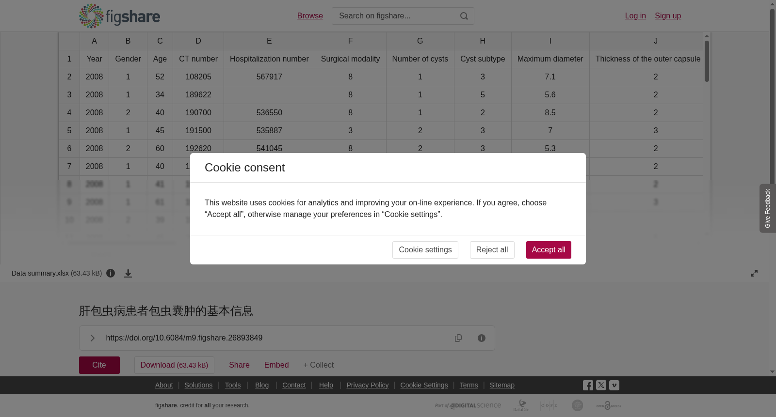
肝包虫病患者包虫囊肿的基本信息
收藏DataCite Commons2024-09-04 更新2024-11-06 收录
下载链接:
https://figshare.com/articles/dataset/__/26893849/2
下载链接
链接失效反馈官方服务:
资源简介:
背景: 本研究探讨了肝棘球蚴成像的 CT 值与收缩胆瘘和钙化等相关参数与肝囊型棘球蚴病 (HCE) 的活动和病程之间的关系。方法: 回顾性分析 331 例经临床 CT 诊断并通过手术病理证实的 HCE 患者的 CT 影像学表现、CT 值和生物学特征。结果: 分期组间囊壁 (F=384.364,P<0.001) 和囊内 (F=217.753,P<0.001) CT 值差异显著,随着 HCE 从 CL 向 CE5 的演变,这两个值都有逐渐增加的趋势。收缩胆瘘的发生率 (χ2=43.902,P<0.001) 和钙化 (χ2=87.488,P<0.001) 的发生率随着 HCE 从 CL 到 CE5 的演变而逐渐增加;CT 值与钙化 (rs=0.468) 和收缩胆瘘 (rs=0.285) 的发生率呈正相关;收缩胆瘘与钙化呈正相关 (χ2=6.972,P=0.008)。CE1 (单房) 和 CE2 (多房) 分期的囊内 CT 值分别为 11.84±6.23 (胡) 和 19.65±8.46 (胡),囊壁值为 32.36。±8.36 (胡) 和 46.52±10.78 (胡)。CE3 (内部囊膜塌陷)、CE4 (实变) 和 CE5 (钙化) 期囊壁的 CT 值分别为 65.23±17.45 (胡) 、 97.345±32.32 (胡) 和 192.23±52.94 (胡)。CE3 、 CE4 和 CE5 阶段的囊内值分别为 31.56±10.89 (胡) 、 43.85±11.23 (胡) 和 95.36±43.89 (胡)。结论: CT值和结果可以反映肝棘球菌不同阶段的囊肿含量和钙化趋势,对判断肝棘球菌胆瘘的发生和钙化的发生以及预测包虫的生长状态具有一定的价值。在 HCE 中,由于收缩胆瘘、钙化和感染导致生物活性降低,以及通过慢速和快速途径,包虫死亡逐渐发生。结果支持囊性肝棘球菌感染是一种缓慢死亡途径的理论。
Background: This study investigated the correlation between CT values of hepatic echinococcosis imaging, related parameters including contracted biliary fistula and calcification, and the activity and disease course of hepatic cystic echinococcosis (HCE).
Methods: A retrospective analysis was conducted on 331 HCE patients who were clinically diagnosed by CT and confirmed via surgical pathology, with their CT imaging findings, CT values and biological features analyzed.
Results: Significant differences in CT values were observed between the staging groups for both the cyst wall (F=384.364, P<0.001) and intracystic contents (F=217.753, P<0.001). As HCE evolved from CL to CE5, both CT values showed a gradual upward trend. The incidences of contracted biliary fistula (χ²=43.902, P<0.001) and calcification (χ²=87.488, P<0.001) increased progressively as HCE progressed from CL to CE5. CT values were positively correlated with the incidence of calcification (rs=0.468) and contracted biliary fistula (rs=0.285); contracted biliary fistula was positively correlated with calcification (χ²=6.972, P=0.008). The intracystic CT values of CE1 (unilocular) and CE2 (multilocular) stages were 11.84±6.23 (HU) and 19.65±8.46 (HU), respectively, while their cyst wall CT values were 32.36±8.36 (HU) and 46.52±10.78 (HU), respectively. The cyst wall CT values of CE3 (internal cyst membrane collapse), CE4 (consolidation) and CE5 (calcification) stages were 65.23±17.45 (HU), 97.345±32.32 (HU) and 192.23±52.94 (HU), respectively. The intracystic CT values of CE3, CE4 and CE5 stages were 31.56±10.89 (HU), 43.85±11.23 (HU) and 95.36±43.89 (HU), respectively.
Conclusions: CT values and related parameters can reflect the cystic contents and calcification trends of HCE cysts at different stages, and possess certain clinical value for judging the occurrence of biliary fistula and calcification in HCE, as well as predicting the growth status of echinococcus. In HCE, the death of echinococcus gradually occurs due to reduced biological activity caused by contracted biliary fistula, calcification and infection, through both slow and rapid pathways. The results support the theory that cystic hepatic echinococcosis follows a slow death pathway.
提供机构:
figshare
创建时间:
2024-09-04
搜集汇总
数据集介绍
背景与挑战
背景概述
该数据集包含331例肝囊型棘球蚴病患者的CT影像和生物学特征数据,重点研究不同疾病分期的CT值变化及其与胆瘘、钙化的相关性,为判断疾病活动状态提供临床依据。
以上内容由遇见数据集搜集并总结生成


