public-4dstem
收藏资源简介:
ECLIPSE public 4D-STEM 是一个公开的4D扫描透射电子显微镜(4D-STEM)数据集集合。它汇集了来自其他研究组已发表的实验4D-STEM数据,并以统一的、无损压缩的HDF5格式(扩展名为.e4d)重新托管,且带有完整且经过验证的校准元数据。该数据集旨在为各种剂量范围内的重建、压缩和去噪方法提供基准测试平台,涵盖叠层衍射(ptychography)和纳米束衍射(nanobeam diffraction)两种模态。每个文件均保证无损性(原始数据与声明变换后的立方体完全一致)、校准完整性(能量、汇聚半角、扫描步长、探测器采样、扫描-探测器旋转、离焦量等参数均标明来源并经校验)以及剂量信息(标明是电子计数、原始探测器单位还是预处理值)。数据集中包含一个固定的基准感兴趣区域(benchmark_roi),便于进行可比较的基准测试。目录表列出了19个数据集,涉及多个材料体系(如STO、NNO、La3Ni2O7、PdCuSi、polySi、PdH、SiGe、NVdiamond、AlAu、UCNP、SAEP、PBTTT、AlSPED、MOF、apoF、MAPbI3、MoS2MoSe2、Sb2S3),剂量范围从约6 e/Ų到5.7e6 e/Ų,探测器包括Merlin Medipix3RX、EMPAD、Gatan K3、Dectris ELA、4D Camera、MerlinEM、Gatan CCD、Timepix3等,加速电压从80 kV到300 kV不等。数据文件按scan_y, scan_x, k_y, k_x的四维立方体存储,按扫描行分块,使用Blosc2 zstd + bitshuffle压缩。每个数据集可能有多个变体(如不同区域或实验条件)。所有数据集均要求引用原始出版物,本仓库仅作为统一格式的重新托管平台。
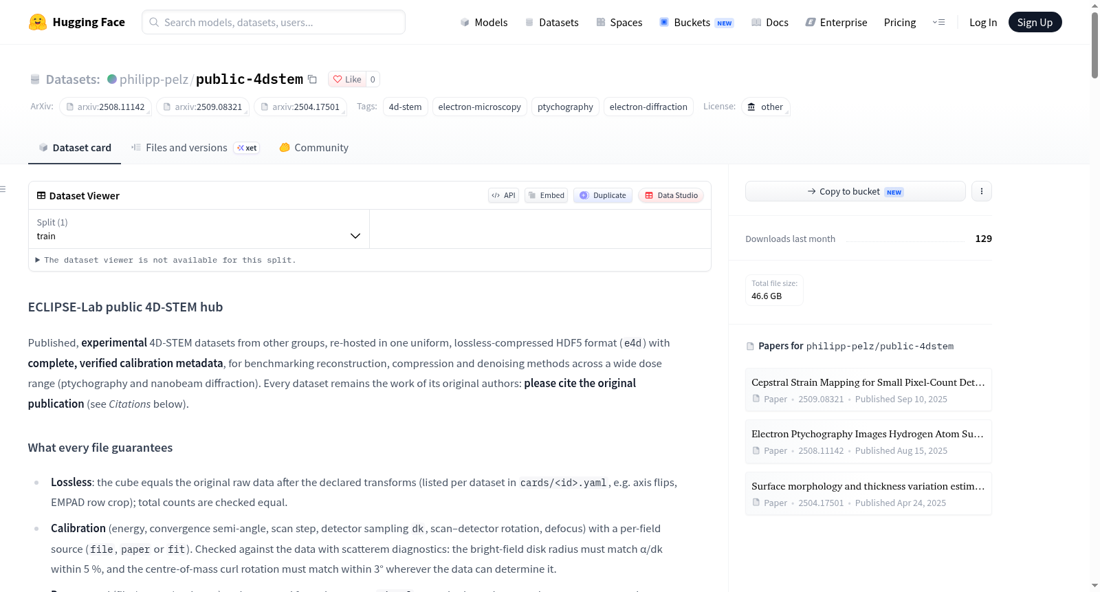
ECLIPSE public 4D-STEM is a publicly available collection of 4D scanning transmission electron microscopy (4D-STEM) datasets. It compiles published experimental 4D-STEM data from other research groups and redistributes them in a unified, losslessly compressed HDF5 format (extension .e4d) with complete and validated calibration metadata. This dataset aims to provide a benchmark platform for reconstruction, compression, and denoising methods across various dose regimes, covering both ptychography and nanobeam diffraction modalities. Each file guarantees losslessness (the raw data is identical to the declared transformed cube), calibration completeness (energy, convergence semi-angle, scan step, detector sampling, scan-detector rotation, defocus, etc., are sourced and verified), and dose information (indicating whether it is electron counts, raw detector units, or preprocessed values). The dataset includes a fixed benchmark region of interest (benchmark_roi) to enable comparable benchmarks. The table of contents lists 19 datasets, covering multiple material systems (e.g., STO, NNO, La3Ni2O7, PdCuSi, polySi, PdH, SiGe, NVdiamond, AlAu, UCNP, SAEP, PBTTT, AlSPED, MOF, apoF, MAPbI3, MoS2MoSe2, Sb2S3), with dose ranges from approximately 6 e/Ų to 5.7e6 e/Ų, detectors including Merlin Medipix3RX, EMPAD, Gatan K3, Dectris ELA, 4D Camera, MerlinEM, Gatan CCD, Timepix3, etc., and accelerating voltages from 80 kV to 300 kV. Data files are stored as four-dimensional cubes (scan_y, scan_x, k_y, k_x), chunked along scan rows, and compressed using Blosc2 zstd + bitshuffle. Each dataset may have multiple variants (e.g., different regions or experimental conditions). All datasets require citation of the original publications; this repository serves only as a redistribution platform in a unified format.
ECLIPSE public 4D-STEM 数据集
数据集概述
- 名称:ECLIPSE public 4D-STEM
- 许可:other
- 标签:4d-stem、electron-microscopy、ptychography、electron-diffraction
- 内容:来自其他课题组的已发表实验性 4D-STEM 数据集,统一重托管为无损压缩的 HDF5 格式(
e4d),包含完整且经验证的校准元数据,用于在宽剂量范围内(ptychography 与 nanobeam diffraction)对重建、压缩和去噪方法进行基准测试。 - 版权说明:每个数据集仍归原作者所有,使用时请引用原始出版物。
每个文件的保证
无损性
- 数据立方体等于原始原始数据在声明变换(如轴翻转、EMPAD 行裁剪,按数据集列于
cards/<id>.yaml)之后的结果;总计数经检查相等。
校准
- 包括能量、会聚半角、扫描步长、探测器采样
dk、扫描–探测器旋转、离焦,每项均标注来源(file、paper或fit)。 - 通过 scatterem 诊断对数据进行检查:明场盘半径须与 α/dk 在 5% 内匹配,在数据可判定的情况下质心旋度旋转须在 3° 内匹配。
剂量
- 声明剂量(来自 file / paper / estimate)并从计数中测量。
signal字段说明数值是电子计数、原始探测器单位(ADU)还是预处理(归一化)值。
基准 ROI
- 每个数据集具有固定的
benchmark_roi,用于可比较的基准测试。
数据集目录
| id | modality | tasks | variants | 声明剂量 (e/Ų) | 测量剂量 (e/Ų) | signal | detector | kV | licence | GB | original DOI |
|---|---|---|---|---|---|---|---|---|---|---|---|
| Strauch2021_STO | ptycho | ptycho | 1 | 5.7e+06 (paper) | 5.72e+06 | counts | Merlin Medipix3RX | 300 | CC-BY-4.0 | 0.13 | 10.1017/S1431927621012423 |
| Harikrishnan2025_NNO | ptycho | ptycho | 1 | 2.2e+06 (paper) | 2.01e+06 | counts | EMPAD | 300 | CC-BY-4.0 | 0.88 | 10.5281/zenodo.14954707 |
| Dong2024_La3Ni2O7 | ptycho | ptycho | 3 | 9e+05 (estimate) | 7.49e+05–7.87e+05 | counts | Gatan K3 (energy-filtered) | 300 | CC-BY-4.0 | 0.57 | 10.1038/s41586-024-07482-1 |
| Riechers2026_PdCuSi | nbed | amorphous | 2 | 3e+05 (estimate) | 6.38e+05–7.28e+05 | counts | Dectris ELA | 200 | CC-BY-4.0 | 2.87 | 10.1016/j.jallcom.2026.186631 |
| Kang2025_polySi | ptycho | ptycho | 1 | 7.5e+05 (estimate) | 5.68e+05 | counts | Gatan K3 | 300 | CC-BY-4.0 | 0.39 | 10.26599/NR.2025.94907398 |
| Shi2025_PdH | ptycho | ptycho | 3 | 2e+05–5e+05 (estimate/file) | 2.07e+05–5.28e+05 | adu | EMPAD | 300 | CC-BY-4.0 | 7.42 | 10.48550/arXiv.2508.11142 |
| Sadri2024_STO | ptycho | ptycho, virtual_imaging | 2 | 6.39–3.5e+05 (paper) | 6.35–3.7e+05 | counts | EMPAD | 300 | CC0-1.0 | 0.58 | 10.1038/s41524-024-01428-x |
| KP2025_cepstral_SiGe | nbed | strain, virtual_imaging | 6 | 6.24e+03–6.24e+04 (estimate) | – | adu | EMPAD | 300 | CC-BY-4.0 | 4.74 | 10.48550/arXiv.2509.08321 |
| Chen2025_NVdiamond | ptycho | ptycho | 2 | 2e+05 (paper) | 1.25e+05–1.33e+05 | counts | Gatan K3 (energy-filtered) | 300 | CC-BY-4.0 | 0.82 | 10.1016/j.xinn.2025.101043 |
| MillsZeltmann2022_AlAu | nbed | strain, virtual_imaging | 1 | 6.71e+04 (estimate) | – | adu | Gatan K2-IS | 300 | CC-BY-4.0 | 0.08 | 10.1016/j.actamat.2023.118721 |
| Ribet2024_UCNP | ptycho | ptycho | 2 | 5e+03 (paper) | 4.27e+03–4.98e+03 | counts | 4D Camera | 300 | CC-BY-4.0 | 0.51 | 10.1063/5.0207212 |
| Zhang2025_SAEP | ptycho | ptycho | 3 | 44–1.2e+03 (estimate/paper) | 111–2.97e+03 | counts | MerlinEM | 300 | CC-BY-4.0 | 0.26 | 10.48550/arXiv.2504.17501 |
| Balhorn2022_PBTTT | nbed | virtual_imaging | 1 | 156 (file) | – | adu | Gatan CCD (TitanX / NCEM) | 300 | CC-BY-4.0 | 0.91 | 10.1073/pnas.2204346119 |
| Thronsen2022_AlSPED | sped | phase_map, acom | 1 | 1e+03 (estimate) | – | normalized | MerlinEM 1S (Quantum Detectors) | 200 | CC-BY-4.0 | 4.02 | 10.1016/j.ultramic.2023.113861 |
| Li2025_MOF | ptycho | ptycho | 3 | 100 (paper) | 98.9–116 | adu | EMPAD | 300 | CC-BY-4.0 | 11.93 | 10.1038/s41467-025-55827-9 |
| Kucukoglu2024_apoF | ptycho | ptycho | 3 | 34 (paper) | – | adu | Dectris ELA | 300 | CC0-1.0 | 4.35 | 10.1101/2024.02.12.579607 |
| Yuan2025_MAPbI3 | ptycho | ptycho | 3 | 11 (paper) | 11–11 | counts | Timepix3 | 200 | CC-BY-4.0 | 0.03 | 10.1038/s41586-025-09693-6 |
| Mireles2025_MoS2MoSe2 | nbed | strain, virtual_imaging | 1 | 9.94 (paper) | 9.68 | adu | EMPAD | 80 | CC-BY-4.0 | 3.70 | 10.1126/sciadv.adz7908 |
| Wu2025_Sb2S3 | nbed | acom, virtual_imaging | 2 | 6.23 (estimate) | 2.63–2.64 | counts | Dectris Quadro (TESCAN Tensor) | 100 | CC-BY-4.0 | 2.42 | 10.1021/acsnano.5c04342 |
目录布局
data/<id>/<id>[_<variant>].h5— 数据立方体,形状(scan_y, scan_x, k_y, k_x),按扫描行分块,Blosc2 zstd + bitshuffle(需要hdf5plugin)。大于 45 GB 的文件沿scan_y拆分为.part-NN.h5。cards/<id>.yaml— 完整元数据卡片(同时以/metadata.attrs[card_json]嵌入每个文件)。reports/<id>/— 验证报告(JSON)和预览(平均图案、virtual BF / ADF)。
加载方式
使用纯 h5py 加载
python import json, h5py, hdf5plugin with h5py.File(Strauch2021_STO.h5, r) as f: roi = f[data][32:96, 32:96] # 仅读取这些块 card = json.loads(f[metadata].attrs[card_json])
使用 scatterem 加载
python from scatterem.data.public.hub import Hub4DStem, list_datasets ptycho_low_dose = list_datasets(task=ptycho, dose=(1, 1e3)) ds = Hub4DStem(Strauch2021_STO, roi=benchmark) # Dataset4DStem
变体列表
- Harikrishnan2025_NNO:
fig_4b - Dong2024_La3Ni2O7:
region01、region02、region03 - Riechers2026_PdCuSi:
FQ_locC_scan2、SQ_locA_scan3 - Shi2025_PdH:
challenge、non_superlattice_500k、superlattice_200k - Sadri2024_STO:
highmag_hd、lowmag_highangle_hd - KP2025_cepstral_SiGe:
a0p75_1ms、a1p6_1ms、a1p6_10ms、a2_10ms、a3_1ms、a3_10ms - Chen2025_NVdiamond:
fig_3、fig_4 - MillsZeltmann2022_AlAu:
au_beforeHT - Ribet2024_UCNP:
pristine、defect - Zhang2025_SAEP:
beta、mil101、uio66_first - Li2025_MOF:
fig2_zrbtb、fig3_moss6、fig4_moss6 - Kucukoglu2024_apoF:
pos_16、pos_40、pos_51 - Yuan2025_MAPbI3:
fig3_scan0、fig3_scan1、fig3_scan2 - Wu2025_Sb2S3:
prec0、prec1
引用信息(请引用原始工作)
Strauch2021_STO
Strauch et al., Live processing of momentum-resolved STEM data for first moment imaging and ptychography, Microsc. Microanal. (2021) DOI: 10.1017/S1431927621012423 · licence: CC-BY-4.0 · original data: https://zenodo.org/records/5113449
Harikrishnan2025_NNO
Harikrishnan et al., Zenodo 14954707 (NNO multislice ptychography datasets) DOI: 10.5281/zenodo.14954707 · licence: CC-BY-4.0 · original data: https://zenodo.org/records/14954707
Dong2024_La3Ni2O7
Dong et al., Visualization of oxygen vacancies and self-doped ligand holes in La3Ni2O7-δ, Nature 630, 847–852 (2024) DOI: 10.1038/s41586-024-07482-1 · licence: CC-BY-4.0 · original data: https://zenodo.org/records/12807652
Riechers2026_PdCuSi
Riechers et al., Spatial distribution and connectivity of medium-range order signatures in a metallic glass probed with simulated and experimental 4DSTEM, J. Alloys Compd. (2026) DOI: 10.1016/j.jallcom.2026.186631 · licence: CC-BY-4.0 · original data: https://zenodo.org/records/18549114
Kang2025_polySi
Kang et al., Thermal cycle impact on polycrystalline silicon: Direct observation of electrical properties degradation and interfacial nanocrystalline grain defects, Nano Research (2025) DOI: 10.26599/NR.2025.94907398 · licence: CC-BY-4.0 · original data: https://zenodo.org/records/15089477
Shi2025_PdH
Shi et al., Electron Ptychography Images Hydrogen Atom Superlattices and 3D Inhomogeneities in Palladium Hydride Nanoparticles, arXiv:2508.11142 (2025) DOI: 10.48550/arXiv.2508.11142 · licence: CC-BY-4.0 · original data: https://zenodo.org/records/21363131
Sadri2024_STO
Sadri et al., Unsupervised deep denoising for four-dimensional scanning transmission electron microscopy, npj Comput. Mater. 10, 243 (2024) DOI: 10.1038/s41524-024-01428-x · licence: CC0-1.0 · original data: https://doi.org/10.6084/m9.figshare.25815436
KP2025_cepstral_SiGe
Harikrishnan KP et al., Cepstral Strain Mapping for Small Pixel-Count Detectors, arXiv:2509.08321 (2025) DOI: 10.48550/arXiv.2509.08321 · licence: CC-BY-4.0 · original data: https://zenodo.org/records/18065811
Chen2025_NVdiamond
Chen et al., Visualizing the Atomic Structure of Nitrogen-vacancy Color Center in Diamond by Multislice Electron Ptychography, The Innovation 7, 101043 (2025) DOI: 10.1016/j.xinn.2025.101043 · licence: CC-BY-4.0 · original data: https://zenodo.org/records/14913471
MillsZeltmann2022_AlAu
Mills et al., Nanoscale mapping of point defect concentrations with 4D-STEM, Acta Materialia (2023) DOI: 10.1016/j.actamat.2023.118721 · licence: CC-BY-4.0 · original data: https://zenodo.org/records/7041997
Ribet2024_UCNP
Ribet et al., Uncovering the three-dimensional structure of upconverting core–shell nanoparticles with multislice electron ptychography, Appl. Phys. Lett. (2024) DOI: 10.1063/5.0207212 · licence: CC-BY-4.0 · original data: https://zenodo.org/records/10775819
Zhang2025_SAEP
Zhang et al., Surface morphology and thickness variation estimation of zeolites via electron ptychography, arXiv:2504.17501 (2025) DOI: 10.48550/arXiv.2504.17501 · licence: CC-BY-4.0 · original data: https://zenodo.org/records/17709147
Balhorn2022_PBTTT
Balhorn et al., Closing the loop between microstructure and charge transport in conjugated polymers by combining microscopy and simulation, Proc. Natl. Acad. Sci. U.S.A. 119, e2204346119 (2022) DOI: 10.1073/pnas.2204346119 · licence: CC-BY-4.0 · original data: https://zenodo.org/records/6585396
Thronsen2022_AlSPED
Thronsen et al., Scanning precession electron diffraction data analysis approaches for phase mapping of precipitates in aluminium alloys, Ultramicroscopy (2023) DOI: 10.1016/j.ultramic.2023.113861 · licence: CC-BY-4.0 · original data: https://zenodo.org/records/6645396
Li2025_MOF
Li et al., Atomically resolved imaging of radiation-sensitive metal-organic frameworks via electron ptychography, Nat. Commun. 16 (2025) DOI: 10.1038/s41467-025-55827-9 · licence: CC-BY-4.0 · original data: https://zenodo.org/records/13958144
Kucukoglu2024_apoF
Küçükoğlu et al., Low-dose cryo-electron ptychography of proteins at sub-nanometer resolution, bioRxiv (2024) DOI: 10.1101/2024.02.12.579607 · licence: CC0-1.0 · original data: https://www.ebi.ac.uk/empiar/EMPIAR-12236/
Yuan2025_MAPbI3
Yuan et al., Atomically resolved edges and defects in lead halide perovskites, Nature 647, 364–368 (2025) DOI: 10.1038/s41586-025-09693-6 · licence: CC-BY-4.0 · original data: https://zenodo.org/records/11482208
Mireles2025_MoS2MoSe2
Mireles et al., Strain mapping of three-dimensionally structured two-dimensional materials, Sci. Adv. (2026) DOI: 10.1126/sciadv.adz7908 · licence: CC-BY-4.0 · original data: https://zenodo.org/records/17246822
Wu2025_Sb2S3
Wu et al., Correlative and in situ microscopy investigation of phase transformation, crystal growth, and degradation of antimony sulfide thin films, ACS Nano (2025) DOI: 10.1021/acsnano.5c04342 · licence: CC-BY-4.0 · original data: https://zenodo.org/records/15536234