fNIRS dataset for the analysis of oxygen saturation in the primary motor cortex during a single hand movement.
收藏资源简介:
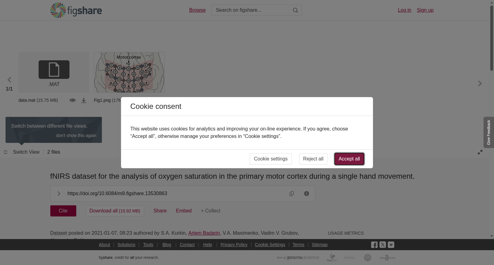
Sixteen healthy volunteers (age: 20-45 years, males) participated in the experiment. All the participants were right-handed, amateur practitioners of physical exercises, and non-smokers. The subjects were conditionally healthy: no diagnosed diseases of the musculoskeletal or nervous system, no prescribed medications. Prior to the experiment, the participants were asked to maintain a healthy lifestyle for a least 48 hours, including 8-hours night rest, prohibited alcohol consumption, limited caffeine consumption, and moderate physical activity.The experiments were held in the first half of the day in a room with a sufficient natural lighting level. Before the beginning of the experiment, the volunteers were instructed about its goals and methods, along with possible inconveniences. The participants were able to ask any related questions and receive detailed answers. After all explanations, each participant filled and signed the blank of informed written consent before participating in the experiment. The experimental procedure was performed following the Helsinki’s Declaration and approved by the local Ethics Committee of the Innopolis University.The fNIRS signals were recorded with the “NIRScout” device by “NIRx” (Germany). The NIRScout system uses 8 sources and 8 detectors to record hemodynamic response in the cortex with a sampling rate of 7.8125 Hz. Optodes are placed non-invasively on the subject’s scalp by mounting to the sockets of the special cap by “EASYCAP” (Germany). FNIRS channels were formed by “source-detector” pairs that were placed close enough to each other. The optimal distance between source and detector is about 3 cm due to the specific traveling path of near-infrared light in cortex’s tissue: the light diffuses in the tissue and at a depth of approximately 3 cm is reflected towards the detector, forming a so-called “banana-shaped” path.In our experiments, we placed optodes in the primary motor cortex area (M1), as shown in Fig. 1. Fig. 1 illustrates our montage with overall 23 fNIRS channels. Additionally, 8 EEG electrodes located according to the international scheme “10-10” are shown as the reference for a better understanding of optodes’ placements.EMG signals were recorded with POLY channels of the encephalograph “Encephalan-EEGR-19/26” by “Medicom- MTD” (Russia). In our study, we used special EMG sensors with two recording electrodes and one ground electrode. Such a scheme allows recording the envelope of EMG signal with a sampling rate of 250 Hz. EMG envelope is related to muscular contractions, and in our experiment, it was used to assess hand movements while high-frequency EMG component was filtered (with a low-pass filter with cutoff frequency – 1 Hz). EMG electrodes were placed non-invasively on the forearm muscle on each of the subject’s hands. Before electrode mounting, the skin was treated with abrasive gel “NuPrep” to ensure good conductivity and high quality of recorded EMG signals. As recording electrodes, we used disposable, pre-gelled adhesive electrodes “Sticktrode” by “Neuroelectrics” (Spain).All experimental work was carried out in the Neuroscience and Cognitive Technology Laboratory of the Innopolis University.The experiment was performed as follows. The subject was sitting in a comfortable chair with hands placed on armrests. The monitor was placed in front of the subject’s eyes at a distance of 70-80 cm for demonstration of visual commands. Each experiment began and ended with a 3-min recording of background brain activity, during which the subject was instructed to relax and make no hand movements. During the active phase of the experiment, the subject was asked to perform movements with his/her left or right hand according to the screen instructions. There were 40 total hand movement trials (20 for each hand) that were performed in random order.<br><br>Each hand movement trial consisted of several phases accompanied by specific commands on the monitor. The trial started with the fixation of the subject’s attention — a bright cross appeared on a black screen for 2 seconds and acted as a signal for the subject to prepare for the trial. Attention fixation was followed by a visual cue: the cross stayed on the screen, and the left- or right-oriented arrow appeared on the top of it for 1.5 seconds. During this phase, the subject was informed that the left- or right-hand movement (correspondingly) was required. The next phase was motor execution: the arrow disappeared from the screen, but the cross stayed for 5 seconds. During this time interval, the subject performed the required hand movement. Hand movement consisted of bending and unbending of fingers to the palm’s center, similar to clenching of the imaginary ball. The trial ended with rest: the cross disappeared, and the black screen was shown for 15 seconds. During this phase, the subject rested and waited for the next command.We applied a band-pass filter (0.01 − 0.1 Hz) to pre-process experimental fNIRS data before all calculations based on the modified Beer-Lambert law. FNIRS data acquisition and pre-processing procedures were performed with “NIRScout” software.For each subject, EMG and fNIRS trials were time-locked to the command and averaged across all trials corresponding to the left and right-hand movements. Thus, event-related EMG/fNIRS signals were obtained. On the group level, the 7-s EMG trials were tested against the baseline separately for the left and right hands. The 18-s fNIRS trials were corrected to the baseline as (SaO2 − SaO2base)/SaO2base and contrasted between the left-hand and right-hand movements.
16名健康男性志愿者(年龄20-45岁)参与本实验。所有受试者均为右利手,为业余体育锻炼爱好者且无吸烟史。受试者均为健康状态:无肌肉骨骼或神经系统确诊疾病,未服用任何处方药物。实验前,要求受试者保持健康生活方式至少48小时,包括8小时夜间睡眠、禁止饮酒、限制咖啡因摄入以及适度体育活动。 实验时段均为当日上午,在自然光充足的房间内进行。实验开始前,向志愿者说明实验目的、方法及可能存在的不适,受试者可提出相关疑问并获得详细解答。完成所有说明后,每名受试者需签署书面知情同意书后方可参与实验。本实验流程符合《赫尔辛基宣言》,并经因诺波利斯大学当地伦理委员会批准。 功能近红外光谱(functional near-infrared spectroscopy, fNIRS)信号由德国NIRx公司的"NIRScout"设备采集。NIRScout系统配备8个光源与8个探测器,以7.8125 Hz的采样率记录皮层血液动力学响应。光极(optodes)通过德国EASYCAP公司的专用帽体插槽无创固定于受试者头皮。fNIRS通道由间距足够近的"光源-探测器"对构成。由于近红外光在皮层组织中的特定传播路径,光源与探测器的最佳间距约为3 cm:光线在组织内散射,在约3 cm深度处反射回探测器,形成所谓的"香蕉形"传播路径。 本实验中将光极放置于初级运动皮层(primary motor cortex, M1)区域,如图1所示。图1展示了本实验的电极排布,共包含23个fNIRS通道。此外,图中还标注了按照国际"10-10"系统放置的8个脑电(electroencephalogram, EEG)电极,用于辅助理解光极的摆放位置。 肌电(electromyography, EMG)信号由俄罗斯Medicom-MTD公司的"Encephalan-EEGR-19/26"脑电图仪的POLY通道采集。本研究使用配备2个记录电极与1个接地电极的专用EMG传感器,该配置可采集肌电信号包络,采样率为250 Hz。肌电信号包络与肌肉收缩相关,本实验中通过对高频EMG成分进行滤波(采用截止频率为1 Hz的低通滤波器),以评估手部运动情况。EMG电极无创固定于受试者双侧前臂肌肉。电极安装前,使用"NuPrep"磨砂凝胶处理皮肤,以确保良好的导电性与优质的EMG信号采集质量。记录电极采用西班牙Neuroelectrics公司生产的"Sticktrode"预凝胶一次性粘贴电极。 所有实验工作均在因诺波利斯大学神经科学与认知技术实验室完成。 实验流程如下:受试者坐在舒适的座椅上,双手置于扶手之上。显示器位于受试者眼前70-80 cm处,用于呈现视觉指令。每个实验的开始与结束阶段均为3分钟的背景脑活动记录时段,期间要求受试者放松且不得进行手部运动。实验主动阶段,受试者需根据屏幕指令完成左手或右手运动。总计包含40次手部运动试次(每侧手各20次),试次顺序随机排列。 每次手部运动试次包含多个阶段,并伴随屏幕上的特定指令。试次起始为注意力固定期:黑色屏幕上出现亮十字,持续2秒,作为受试者准备试次的信号。注意力固定期过后为视觉提示阶段:十字仍保留在屏幕上,其上方出现指向左或右的箭头,持续1.5秒。此阶段告知受试者需执行对应侧手的运动。下一阶段为运动执行期:箭头消失,十字保留5秒,此时间段内受试者完成指定的手部运动。手部运动为手指向掌心中心弯曲与伸展,类似攥握假想球体的动作。试次最后为休息期:十字消失,屏幕变为黑色,持续15秒,期间受试者休息并等待下一指令。 本研究基于修正的比尔-朗伯定律,在所有计算前对fNIRS实验数据施加带通滤波器(0.01−0.1 Hz)进行预处理。fNIRS数据的采集与预处理流程通过"NIRScout"软件完成。 针对每名受试者,将EMG与fNIRS试次与指令进行时间锁定,并对所有对应左手与右手运动的试次进行平均,从而获得事件相关EMG/fNIRS信号。在组水平分析中,分别针对左手与右手的7秒EMG试次与基线进行对比。18秒的fNIRS试次按照(SaO2 − SaO2base)/SaO2base进行基线校正,并对比左手与右手运动条件下的差异。