High-speed mapping of whole-mouse peripheral nerves at subcellular resolution
收藏DataCite Commons2025-04-27 更新2025-04-16 收录
下载链接:
https://www.scidb.cn/detail?dataSetId=4081aa6d53eb4195907c54c6729a52fe
下载链接
链接失效反馈官方服务:
资源简介:
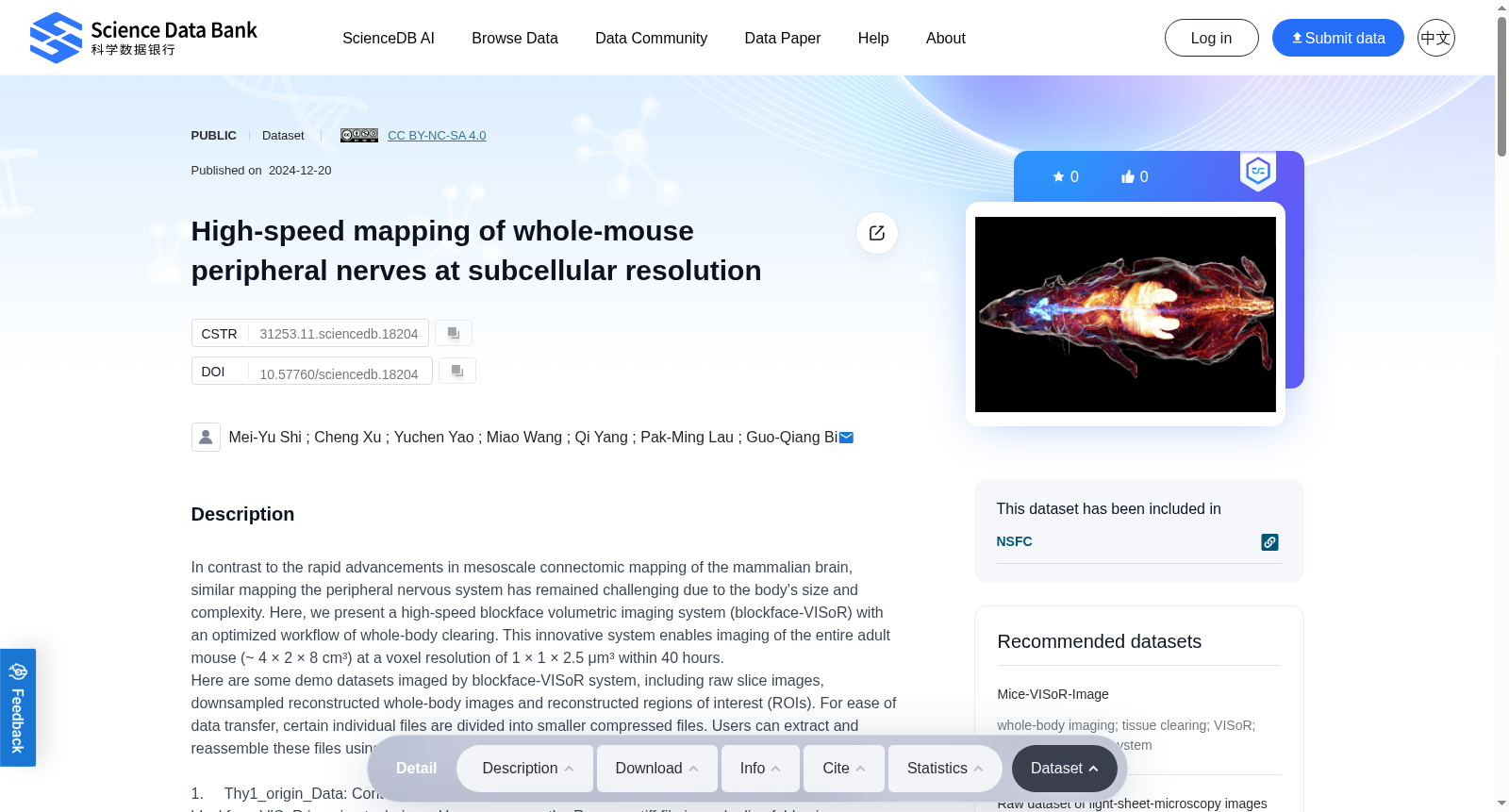
In contrast to the rapid advancements in mesoscale connectomic mapping of the mammalian brain, similar mapping the peripheral nervous system has remained challenging due to the body's size and complexity. Here, we present a high-speed blockface volumetric imaging system (blockface-VISoR) with an optimized workflow of whole-body clearing. This innovative system enables imaging of the entire adult mouse (~ 4 × 2 × 8 cm³) at a voxel resolution of 1 × 1 × 2.5 μm³ within 40 hours.Here are some demo datasets imaged by blockface-VISoR system, including raw slice images, downsampled reconstructed whole-body images and reconstructed regions of interest (ROIs). For ease of data transfer, certain individual files are divided into smaller compressed files. Users can extract and reassemble these files using 7-Zip. 1. Thy1_origin_Data: Contains demo raw volumetric images from a mouse body for validating the blockface-VISoR imaging technique. Users can open the Raw.ome.tiff file in each slice fold using ImageJ/Fiji.2. Thy1_Reconstruction_4μm: Provides 4× downsampled image sequences (at a resolution of 4 × 4 × 4 μm3) of a whole Thy1-EGFP transgenic mouse labeled with tomato lectin conjugated with DyLight 649. The images are compatible with ImageJ/Fiji or can be converted to Imaris format using ImarisFileConverter for viewing in Imaris Viewer or Lychnis.3. Sympathetic ROI: Contains downsampled ROIs of a whole mouse with sympathetic nerves labeling. These .ims format files are viewable in Imaris Viewer or Lychnis.4. Vagal innervations: is an ROI of a whole mouse with vagus nerves labeling. This file can be opened by Imaris Viewer or Lychnis. The complete uncompressed raw image datasets of whole mouse are too large (exceeding 70 terabytes per channel) for public data repositories. A free and more convenient view of the whole-mouse images are publicly accessible through our website (https://smart.siat.ac.cn/meso-example.html). Additionally, other datasets involved in our published work are available to all scientists upon request to the lead contact by feasible data transferring methods including cloud storage, physical hard disk drives, or onsite visiting.
相较于哺乳动物大脑介观尺度连接组图谱绘制的快速进展,外周神经系统的同类图谱绘制仍因机体的体积与复杂性而极具挑战。本研究报道一款搭载优化全身体积透明化流程的高速块面体积成像系统(blockface-VISoR)。该创新系统可在40小时内,以1×1×2.5 μm³的体素分辨率完成整只成年小鼠(约4×2×8 cm³)的成像。
本数据集包含由blockface-VISoR系统成像的演示数据集,涵盖原始切片图像、降采样重建全身体图像以及重建感兴趣区域(ROIs)。为便于数据传输,部分单个文件已拆分为多个压缩子文件,用户可通过7-Zip对其进行解压与重组。
1. Thy1_origin_Data:包含用于验证blockface-VISoR成像技术的小鼠原始体积图像演示数据。用户可通过ImageJ/Fiji打开每个切片文件夹内的Raw.ome.tiff文件。
2. Thy1_Reconstruction_4μm:提供经DyLight 649标记的番茄凝集素染色的Thy1-EGFP转基因整只小鼠的4倍降采样图像序列(分辨率为4×4×4 μm³)。该图像可直接用ImageJ/Fiji打开,或通过ImarisFileConverter转换为Imaris格式后,在Imaris Viewer或Lychnis中查看。
3. Sympathetic ROI:包含经交感神经标记的整只小鼠的降采样感兴趣区域数据。这些.ims格式文件可在Imaris Viewer或Lychnis中打开查看。
4. Vagal innervations:包含经迷走神经标记的整只小鼠的感兴趣区域数据,该文件可通过Imaris Viewer或Lychnis打开。
完整未压缩的小鼠原始图像数据集单通道容量即超过70太字节,无法在公共数据仓库中存储。用户可通过本团队官网(https://smart.siat.ac.cn/meso-example.html)免费便捷地查看整只小鼠的成像结果。此外,本研究已发表工作涉及的其余数据集,可通过联系通讯作者并采用云存储、物理硬盘寄送或现场访问等可行的数据传输方式获取。
提供机构:
Science Data Bank
创建时间:
2024-12-20
搜集汇总
数据集介绍
背景与挑战
背景概述
该数据集提供了小鼠外周神经系统的高分辨率成像数据,包括原始切片图像、下采样重建的全身图像和重建的感兴趣区域(ROIs)。数据集的特点是使用blockface-VISoR系统实现了高分辨率(1 × 1 × 2.5 μm³)和快速成像(40小时内完成整个成年小鼠的成像),支持多种图像处理软件。
以上内容由遇见数据集搜集并总结生成


