
Aerobic Glycolysis Imaging PET-MRI
收藏OpenNeuro2022-03-05 更新2026-03-14 收录
下载链接:
https://openneuro.org/datasets/ds004054
下载链接
链接失效反馈官方服务:
资源简介:
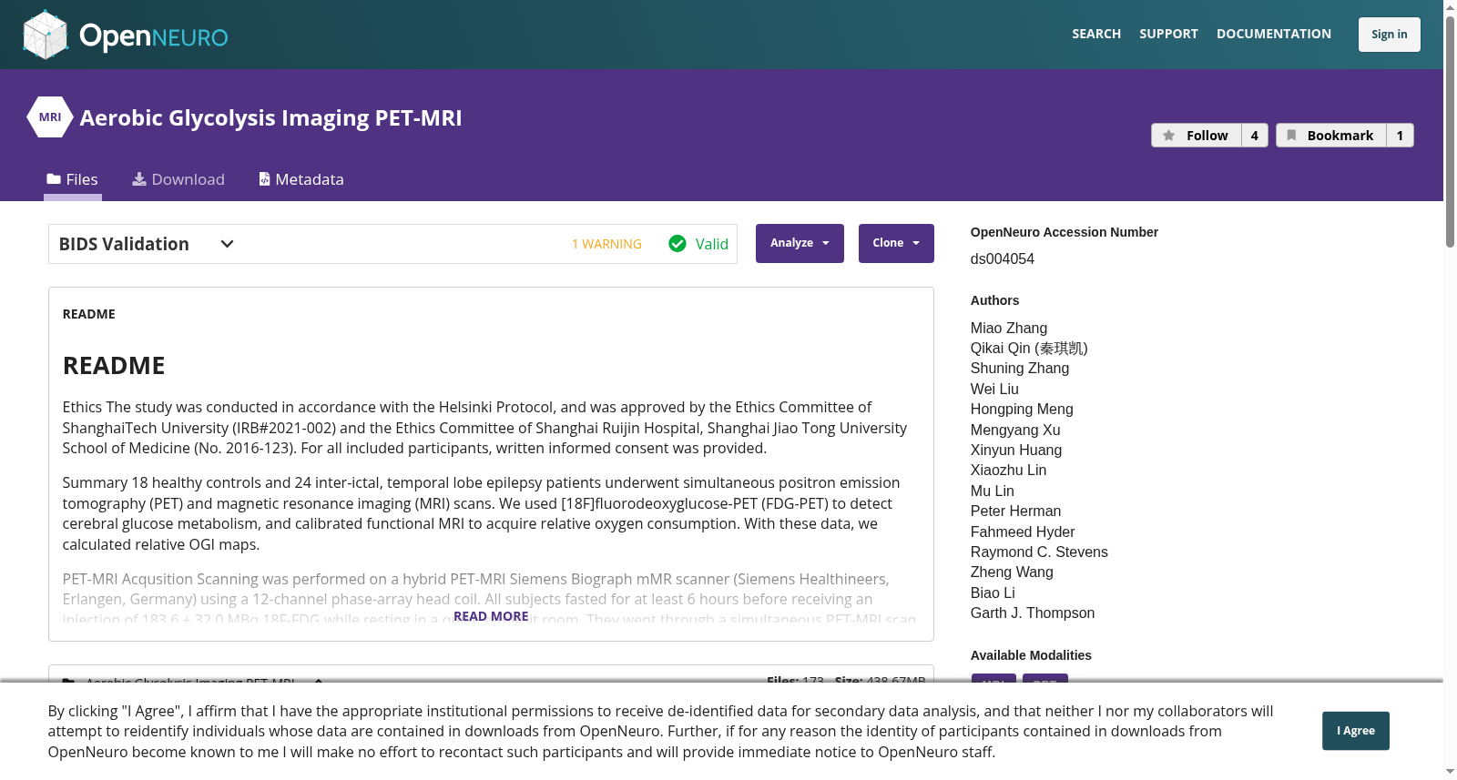
# README
Ethics
The study was conducted in accordance with the Helsinki Protocol, and was approved by the Ethics Committee of ShanghaiTech University (IRB#2021-002) and the Ethics Committee of Shanghai Ruijin Hospital, Shanghai Jiao Tong University School of Medicine (No. 2016-123). For all included participants, written informed consent was provided.
Summary
18 healthy controls and 24 inter-ictal, temporal lobe epilepsy patients underwent simultaneous positron emission tomography (PET) and magnetic resonance imaging (MRI) scans. We used [18F]fluorodeoxyglucose-PET (FDG-PET) to detect cerebral glucose metabolism, and calibrated functional MRI to acquire relative oxygen consumption. With these data, we calculated relative OGI maps.
PET-MRI Acqusition
Scanning was performed on a hybrid PET-MRI Siemens Biograph mMR scanner (Siemens Healthineers, Erlangen, Germany) using a 12-channel phase-array head coil. All subjects fasted for at least 6 hours before receiving an injection of 183.6 ± 32.0 MBq 18F-FDG while resting in a quiet, dimly lit room. They went through a simultaneous PET-MRI scan 40 min later, including static 18F-FDG-PET, sagittal T1-weighted imaging(T1WI), axial Pulsed Arterial Spin Labeling (PASL), axial transverse relaxation time (T2) mapping, and axial observed transverse relaxation time (T2*) mapping. PASL images were converted to quantitative cerebral blood-flow maps with the built-in QUantitative Imaging of Perfusion using a Single Subtraction (QUIPSS) method.16, 17 For T1WI, we used Magnetization Prepared RApid Gradient Echo (MPRAGE) sequence, echo time (TE) = 2.44 ms, repetition time (TR) = 1900 ms, flip angle = 9º. For T2* mapping, we used Gradient Recalled Echo (GRE) sequence: TE = 2.46 / 4.92 / 7.38 / 9.84 ms, TR = 391 ms, flip angle = 25º. For T2 mapping, we used Spin Echo (SE) sequence: TE = 10.5 / 21.0 / 31.5 / 42.0 /52.5 / 63.0 ms, TR = 2000 ms, flip angle = 180º. For axial PASL: TE = 11 ms, TR = 2500 ms, flip angle = 90º.
# README
## 伦理声明
本研究严格遵循赫尔辛基议定书(Helsinki Protocol)开展,并已获得上海科技大学伦理委员会(IRB#2021-002)以及上海交通大学医学院附属瑞金医院伦理委员会(编号:2016-123)的批准。所有纳入研究的受试者均已签署书面知情同意书。
## 研究概述
本研究共纳入18名健康对照者与24名发作间期颞叶癫痫患者,所有受试者均接受了同步正电子发射断层扫描(positron emission tomography, PET)与磁共振成像(magnetic resonance imaging, MRI)检查。本研究采用[18F]氟代脱氧葡萄糖-PET([18F]fluorodeoxyglucose-PET, FDG-PET)检测脑葡萄糖代谢水平,并通过校准功能磁共振成像获取相对氧消耗量。基于上述数据,我们计算得到相对OGI图谱。
## PET-MRI 采集方案
扫描采用西门子Siemens Healthineers公司(德国埃尔朗根)生产的一体化PET-MRI Siemens Biograph mMR扫描仪,并使用12通道相控阵头部线圈。所有受试者在注射183.6±32.0 MBq的18F-FDG前需禁食至少6小时,注射期间需在安静昏暗的房间内静息。注射完成40分钟后,所有受试者接受同步PET-MRI扫描,扫描序列包括静态18F-FDG-PET、矢状位T1加权成像(T1WI)、轴位脉冲动脉自旋标记(Pulsed Arterial Spin Labeling, PASL)、轴位横向弛豫时间(T2)测绘、轴位观测横向弛豫时间(T2*)测绘。采用内置的单次减法定量灌注成像(QUantitative Imaging of Perfusion using a Single Subtraction, QUIPSS)方法,将PASL图像转换为定量脑血流图谱[16, 17]。针对T1加权成像(T1WI),我们采用磁化准备快速梯度回波(Magnetization Prepared RApid Gradient Echo, MPRAGE)序列,其回波时间(echo time, TE)为2.44 ms,重复时间(repetition time, TR)为1900 ms,翻转角为9°。针对T2*测绘,我们采用梯度回波(Gradient Recalled Echo, GRE)序列:回波时间为2.46 / 4.92 / 7.38 / 9.84 ms,重复时间为391 ms,翻转角为25°。针对T2测绘,我们采用自旋回波(Spin Echo, SE)序列:回波时间为10.5 / 21.0 / 31.5 / 42.0 /52.5 / 63.0 ms,重复时间为2000 ms,翻转角为180°。针对轴位PASL:回波时间为11 ms,重复时间为2500 ms,翻转角为90°。
创建时间:
2022-03-05
搜集汇总
数据集介绍
背景与挑战
背景概述
该数据集是一个包含42名参与者(18名健康对照和24名颞叶癫痫患者)的PET-MRI神经影像数据集,用于研究有氧糖酵解成像。数据通过同时采集的18F-FDG-PET和多种MRI序列(包括T1加权、动脉自旋标记、T2和T2*映射)获取,旨在计算相对氧葡萄糖指数(OGI)图以分析脑部葡萄糖代谢和氧消耗。
以上内容由遇见数据集搜集并总结生成


