DRIVE:用于提取血管的数字视网膜图像数据集
收藏帕依提提2024-03-04 收录
下载链接:
https://www.payititi.com/opendatasets/show-26490.html
下载链接
链接失效反馈官方服务:
资源简介:
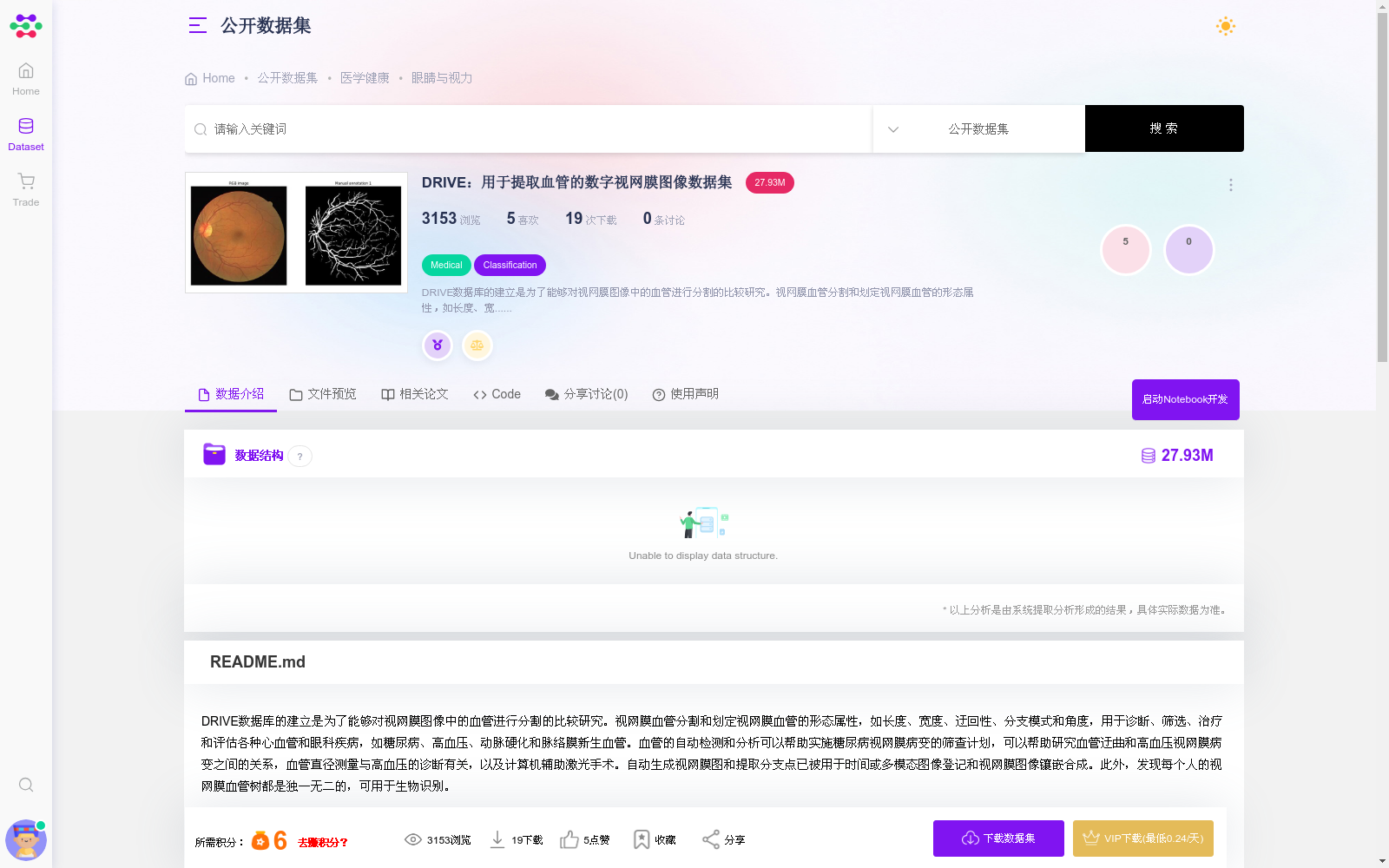
DRIVE数据库的建立是为了能够对视网膜图像中的血管进行分割的比较研究。视网膜血管分割和划定视网膜血管的形态属性,如长度、宽度、迂回性、分支模式和角度,用于诊断、筛选、治疗和评估各种心血管和眼科疾病,如糖尿病、高血压、动脉硬化和脉络膜新生血管。血管的自动检测和分析可以帮助实施糖尿病视网膜病变的筛查计划,可以帮助研究血管迂曲和高血压视网膜病变之间的关系,血管直径测量与高血压的诊断有关,以及计算机辅助激光手术。自动生成视网膜图和提取分支点已被用于时间或多模态图像登记和视网膜图像镶嵌合成。此外,发现每个人的视网膜血管树都是独一无二的,可用于生物识别。 DRIVE数据库的照片来自荷兰的一个糖尿病视网膜病变筛查项目。筛查人群包括400名年龄在25-90岁的糖尿病患者。随机抽取了40张照片,其中33张没有显示任何糖尿病视网膜病变的迹象,7张显示有轻度早期糖尿病视网膜病变的迹象。以下是对这7个病例的异常情况的简要描述。 25_training:色素上皮改变,可能是蝶形黄斑病变伴中央凹色素性瘢痕,或脉络膜病变,无糖尿病视网膜病变或其他血管异常。26_training:背景糖尿病视网膜病变,色素上皮萎缩,视盘周围萎缩 32_training:背景糖尿病视网膜病变 03_test:背景糖尿病视网膜病变 08_test:色素上皮改变,中央凹色素性瘢痕,或脉络膜病变,无糖尿病视网膜病变或其他血管异常14_test:背景糖尿病视网膜病变 17_test:背景糖尿病视网膜病变 每个图像都经过 JPEG 压缩。 这些图像是用佳能CR5非滴胶3CCD相机获得的,具有45度的视野(FOV)。每张图像都是以768×584像素、每色平面8比特拍摄的。每张图像的FOV是圆形的,直径约为540像素。对于这个数据库,图像已经围绕FOV进行了裁剪。对于每张图像,都提供了一个遮罩图像,划定了FOV。 这组 40 张图像分为训练集和测试集,均包含 20 张图像。对于训练图像,可以对脉管系统进行单次手动分割。对于测试用例,有两个手动分段可用;一种用作黄金标准,另一种可用于将计算机生成的分割与独立的人类观察者的分割进行比较。此外,每个视网膜图像都有一个掩模图像,指示感兴趣的区域。所有手动分割脉管系统的人类观察者均由经验丰富的眼科医生指导和培训。他们被要求标记他们至少 70% 确定它们是容器的所有像素。 结果应作为单个 zip 文件提交,其中包含二进制 PNG 文件,其中包含测试集中相应文件的血管预测。文件名应为 [1.png, 2.png, ..., 20.png]。将为测试集中的每个图像计算骰子系数(仅考虑掩码内的像素)。根据最高平均骰子系数对排行榜进行排序。 See http://www.isi.uu.nl/Research/Databases/DRIVE/ for the original web-page on this challenge
The DRIVE database was developed to support comparative studies on vessel segmentation in retinal images. Retinal vessel segmentation and the quantification of morphological features of retinal vessels—including length, width, tortuosity, branching patterns, and branching angles—are critical for the diagnosis, screening, treatment, and evaluation of multiple cardiovascular and ophthalmologic disorders, such as diabetes mellitus, hypertension, arteriosclerosis, and choroidal neovascularization. Automatic detection and analysis of retinal vessels can facilitate the implementation of diabetic retinopathy screening campaigns, enable investigation of the association between vascular tortuosity and hypertensive retinopathy, correlate vascular diameter measurements with hypertension diagnosis, and support computer-assisted laser surgery. Automated generation of retinal maps and extraction of vascular branching points have been applied in temporal or multimodal image registration and retinal image mosaic synthesis. Furthermore, each individual’s retinal vascular tree is unique, making it suitable for biometric identification applications.
The images in the DRIVE database were collected from a Dutch diabetic retinopathy screening program. The screened cohort included 400 diabetic patients aged 25 to 90 years. A random sample of 40 images was selected, with 33 showing no signs of diabetic retinopathy and 7 presenting mild, early-stage diabetic retinopathy. A brief description of the abnormalities in these 7 cases is provided below:
25_training: Pigment epithelial changes, potentially consistent with a butterfly-shaped macular lesion with a central foveal pigmentary scar, or a choroidal lesion, with no diabetic retinopathy or other vascular abnormalities.
26_training: Background diabetic retinopathy with pigment epithelial atrophy and peripapillary atrophy.
32_training: Background diabetic retinopathy.
03_test: Background diabetic retinopathy.
08_test: Pigment epithelial changes, central foveal pigmentary scar, or choroidal lesion, with no diabetic retinopathy or other vascular abnormalities.
14_test: Background diabetic retinopathy.
17_test: Background diabetic retinopathy.
All images were compressed in JPEG format. The images were captured using a Canon CR5 non-mydriatic 3CCD camera with a 45-degree field of view (FOV). Each image has a resolution of 768 × 584 pixels, with 8 bits per color channel. The FOV of each image is circular, with a diameter of approximately 540 pixels. For this database, images have been cropped to the boundaries of the FOV. A binary mask image is provided for each image to delineate the FOV.
This set of 40 images is divided into a training set and a test set, each containing 20 images. For training images, a single manual segmentation of the retinal vasculature is provided. For test images, two manual segmentations are available: one serves as the gold standard, while the other allows for comparison of computer-generated segmentations against those produced by independent human observers. Additionally, a mask image is provided for each retinal image to mark the region of interest (ROI). All human annotators who performed the manual vascular segmentations received guidance and training from an experienced ophthalmologist. They were instructed to label all pixels for which they were at least 70% confident that the pixel represented a vessel.
Submissions should be packaged as a single ZIP file containing binary PNG files with vessel segmentation predictions corresponding to the test set images. The filenames of the submission files must be [1.png, 2.png, ..., 20.png]. The Dice coefficient will be calculated for each test image, with only pixels within the provided mask being considered. The competition leaderboard will be ranked according to the highest average Dice coefficient across the test set.
For the original challenge webpage, see http://www.isi.uu.nl/Research/Databases/DRIVE/
提供机构:
帕依提提
搜集汇总
数据集介绍
背景与挑战
背景概述
DRIVE数据集包含40张糖尿病患者的视网膜图像,用于血管分割研究,支持自动检测和分析血管以辅助诊断和治疗。数据集分为训练和测试集,提供手动分割的脉管系统和遮罩图像,适用于算法开发和性能评估。
以上内容由遇见数据集搜集并总结生成


