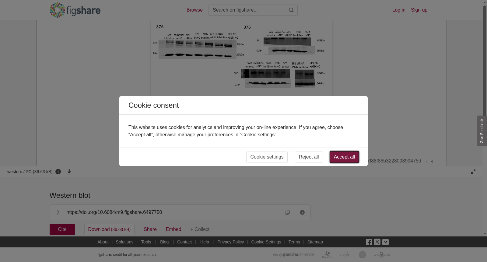
Western blot
收藏资源简介:
Proteins (30μg) extracted from cells were loaded and separated in 8% SDS-PAGE and then transferred on nitrocellulose membranes. The membranes were blocked with 5% non-fat milk and incubated with Anti-SP1 (1:1000, 9389, Cell Signaling Technology, MA, USA) or Anti-CCR7 (1:1000, ab32527, Abcam, Cambridge, UK) in 5% milk/PBS buffer at 4℃ overnight, followed by incubation with goat anti-rabbit secondary antibody (A23720, Abbkine, Wuhan, China) conjugated with DyLight fluorescent 680. After extensive wash with PBST buffer, the band of protein was visualized under Odyssey CLx imaging system (LI-COR, Inc., NE, USA).
从细胞中提取的蛋白质(30μg)经上样后,于8%十二烷基硫酸钠-聚丙烯酰胺凝胶电泳(SDS-PAGE)中完成分离,随后转印至硝酸纤维素膜。以5%脱脂牛奶对膜进行封闭,接着在4℃条件下,用含5%脱脂牛奶的磷酸盐缓冲盐水(PBS)缓冲液配制的抗SP1(Anti-SP1)一抗(稀释比例1:1000,货号9389,美国马萨诸塞州Cell Signaling Technology公司)或抗CCR7(Anti-CCR7)一抗(稀释比例1:1000,货号ab32527,英国剑桥Abcam公司)孵育过夜。随后,使用偶联DyLight荧光染料680的山羊抗兔二抗(货号A23720,中国武汉Abbkine公司)进行孵育。经磷酸盐缓冲盐水-吐温20(PBST)缓冲液充分洗涤后,借助Odyssey CLx成像系统(美国内布拉斯加州LI-COR公司)实现蛋白条带的可视化检测。