
Royal Brisbane_TOFMRA_Intracranial Aneurysm_Database
收藏OpenNeuro2024-04-18 更新2026-03-14 收录
下载链接:
https://openneuro.org/datasets/ds005096
下载链接
链接失效反馈官方服务:
资源简介:
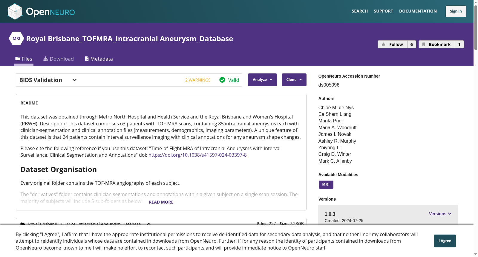
This dataset was obtained through Metro North Hospital and Health Service and the Royal Brisbane and Women's Hospital (RBWH).
Description: This dataset comprises 63 patients with TOF-MRA scans, containing 85 intracranial aneurysms each with clinician-segmentation and clinical annotation files (measurements, demographics, imaging parameters). A unique feature of this dataset is that 24 patients contain interval surveillance imaging with clinical annotations for any aneurysm shape changes.
Please cite the following reference if you use this dataset: "Time-of-Flight MRA of Intracranial Aneurysms with Interval Surveillance, Clinical Segmentation and Annotations" doi: https://doi.org/10.1038/s41597-024-03397-8
## Dataset Organisation
Every original folder contains the TOF-MRA angiography of each subject.
The "derivatives" folder contains clinician segmentations and annotations within a given subject on a single scan session. The majority of subjects will include 5 sub-folders as below:
1) "3D Model": Contains an STL (standard tesselation language) model for the aneurysm-only, the aneurysm with parent artery, and the parent-artery-only. Subjects with multiple aneurysms will have an STL model per aneurysm.
2) "Mask": Contains the nifti angiography with an overlay of the aneurysm mask assigned a pixel value of zero and burnt into the image.
3) "Nifti Aneurysm Only": Contains a nifti file of only the aneurysm segmented mask.
4) "Nifti with Parent Artery": Contains a nifti file of the aneurysm with parent artery segmented mask.
5) "Slicer": Contains the 3D Slicer scene used for segmentation and annotation of the aneurysms and parent arteries.
## Details related to access to the data
- Contact person: Mark C. Allenby (m.allenby@uq.edu.au)
## Overview
- TOF-MRA scans acquired through Metro North Health at various institutions between 2008 and 2022.
- This dataset is intended for research into intracranial aneurysms, and can be used in applications such as the development of automated detection algorithms, in computational fluid dynamics studies, for surgical training models and developing surgical guides, and for evaluating cell behaviour and dynamics in in vitro aneurysm models.
- The supplementary documents provided includes a summary of MR image acquisition parameters for all subjects, patient gender and age, characterises aneurysm location and size, and also provides signal-to-noise (SNR) and contrast-to-noise (CNR) ratios for all scans that were clinically annotated. It might be necessary for users of this dataset to consult the SNR and CNR for patients intended for further research and analysis to ensure accurate segmentation can be achieved.
## Missing Data
Aneurysms in some subjects have been segmentated and annotated using DSA or CTA scans, and thus only STL models have been provided for these subjects as the original files require further de-identification.
本数据集由墨尔本北岸医院与健康服务中心(Metro North Hospital and Health Service)以及皇家布里斯班妇女医院(Royal Brisbane and Women's Hospital,RBWH)联合获取。
## 数据集描述
本数据集包含63名患者的TOF-MRA(Time-of-Flight MRA,时间飞跃法磁共振血管造影)扫描数据,共计85个颅内动脉瘤样本,每份样本均附带临床医师标注的分割结果与临床注释文件(涵盖测量数据、人口统计学信息、成像参数等)。本数据集的独特优势在于,其中24名患者包含间隔随访成像数据,以及针对动脉瘤形态变化的临床注释。
若使用本数据集,请引用以下文献:《Time-of-Flight MRA of Intracranial Aneurysms with Interval Surveillance, Clinical Segmentation and Annotations》,DOI:https://doi.org/10.1038/s41597-024-03397-8
## 数据集组织结构
每个原始文件夹包含对应受试者的TOF-MRA血管造影影像。
"derivatives(衍生数据)"文件夹包含单扫描会话下受试者的临床医师分割结果与注释文件。多数受试者将包含以下5个子文件夹:
1. "3D模型":包含仅动脉瘤、动脉瘤加父动脉,以及仅父动脉的STL(Standard Tessellation Language,标准曲面细分语言)模型;若受试者存在多个动脉瘤,则每个动脉瘤对应一个独立的STL模型。
2. "掩码(Mask)":包含叠加了动脉瘤掩码的NIfTI血管造影影像,掩码像素值设为0并已嵌入图像中。
3. "仅动脉瘤NIfTI文件":仅包含动脉瘤分割掩码的NIfTI文件。
4. "含父动脉的NIfTI文件":包含动脉瘤与父动脉分割掩码的NIfTI文件。
5. "Slicer":包含用于动脉瘤及父动脉分割与注释的3D Slicer场景文件。
## 数据访问相关详情
- 联系人:Mark C. Allenby(电子邮箱:m.allenby@uq.edu.au)
## 数据集概述
- 本数据集的TOF-MRA扫描数据于2008年至2022年间,由墨尔本北岸健康中心旗下多家机构采集。
- 本数据集旨在服务于颅内动脉瘤相关研究,可应用于自动化检测算法开发、计算流体动力学研究、手术训练模型与手术导航工具开发,以及体外动脉瘤模型中的细胞行为与动态特性评估等场景。
- 附带的补充文档涵盖了所有受试者的MR成像采集参数总结、患者性别与年龄信息、动脉瘤位置与大小特征,同时为所有经临床注释的扫描提供了信噪比(SNR)与对比度噪声比(CNR)。本数据集的使用者可能需要查阅对应患者的SNR与CNR数据,以确保后续研究分析中可实现准确的分割操作。
## 数据缺失说明
部分受试者的动脉瘤已通过DSA(Digital Subtraction Angiography,数字减影血管造影)或CTA(CT Angiography,CT血管造影)扫描完成分割与注释,因此仅为这些受试者提供了STL模型;其原始文件仍需进一步去标识化处理。
创建时间:
2024-04-18
搜集汇总
数据集介绍
背景与挑战
背景概述
该数据集包含63名患者的TOF-MRA扫描数据,共85个颅内动脉瘤,每个都有临床医生分割和注释文件,其中24名患者还包含间隔监测成像数据。数据集适用于颅内动脉瘤的多种研究应用,如自动检测算法开发和计算流体动力学研究。
以上内容由遇见数据集搜集并总结生成



