体细胞病理图像分析大模型数据集
收藏资源简介:
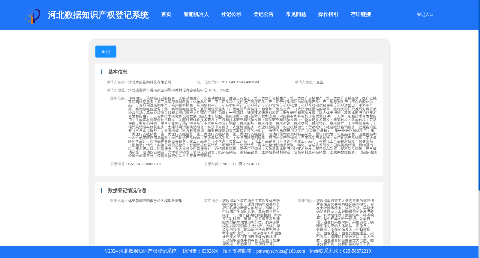
该数据集涵盖了大量高质量的病理切片图像及其关联的临床病理报告。旨在支持肿瘤检测、病变分析、早期疾病筛查以及人工智能模型的开发与验证。具体包括以下数据结构:样本编号:每个样本的唯一标识。采集日期:图像的采集时间。采集部位:病理图像对应的人体部位。图像尺寸、分辨率:图像的像素大小和扫描精度。图像通道:图像的颜色通道。染色方法:病理切片染色方法。放大倍数:图像采集的显微镜放大倍数。图像分析工具:分析图像的软件工具。标注信息:图像的标注内容。病理报告(临床):描述病变特征、诊断意见等文本信息。诊断模型描述:AI 诊断模型的简要描述。诊断模型结果输出:AI 诊断模型的预测结果。准确性:诊断模型输出结果与临床报告一致性的量化评估。
This dataset contains a large number of high-quality pathological slide images and their associated clinical pathology reports. It is designed to support tumor detection, lesion analysis, early disease screening, as well as the development and validation of artificial intelligence models. Specifically, it includes the following data structures: 1. Sample ID: Unique identifier for each individual sample. 2. Collection Date: The timestamp when the image was acquired. 3. Collection Site: The anatomical location of the human body corresponding to the pathological image. 4. Image Size and Resolution: The pixel dimensions and scanning precision of the image. 5. Image Channels: The color channels of the pathological image. 6. Staining Method: The staining protocol used for the pathological slide. 7. Magnification: The microscopic magnification factor utilized during image acquisition. 8. Image Analysis Tool: Software tools employed for image analysis. 9. Annotation Information: The annotated content of the images. 10. Pathological Report (Clinical): Textual information describing lesion features, diagnostic conclusions, and other relevant clinical details. 11. Diagnostic Model Description: A brief overview of the AI-based diagnostic model. 12. Diagnostic Model Output: The prediction results generated by the AI diagnostic model. 13. Accuracy: Quantitative assessment of the consistency between the output results of the diagnostic model and the clinical pathology reports.