基于人工智能的内窥镜图像中仪器分割数据
收藏浙江省数据知识产权登记平台2024-12-16 更新2024-12-17 收录
下载链接:
https://www.zjip.org.cn/home/announce/trends/104921
下载链接
链接失效反馈官方服务:
资源简介:
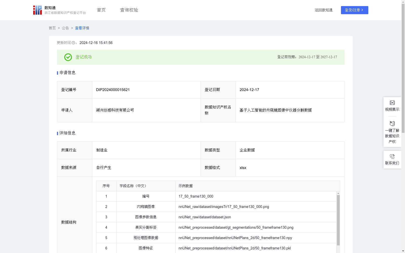
基于人工智能的内窥镜图像中仪器分割技术在微创手术和内窥镜辅助操作中具有重要意义。通过准确分割内窥镜图像中的医疗仪器,系统可以辅助医生在手术中更好地识别和定位器械位置,降低误操作的风险。这项技术在内窥镜引导手术、器械导航和实时操作监控中有广泛应用,有助于提高手术的精确度和安全性,同时减少医生的操作负担。数据收集:在该算法中,首先收集内窥镜图像及其对应的真实分割标签作为训练和验证数据集的基础。每个图像样本包含:内窥镜图像(.png格式文件)和图像参数信息(.json格式),用于记录图像拍摄的参数,帮助模型更好地理解图像特征。此外,真实分割标签标识出仪器的具体位置和轮廓,作为监督学习的目标数据。
数据预处理:对原始内窥镜图像进行预处理,包括缩放、归一化等步骤,以使图像适配神经网络的输入要求。生成的预处理图像数据(.npy格式)和图像特征(.pkl格式)包含了图像的结构化信息,便于模型在细节层面上进行分割。
模型构建:利用基于2D卷积神经网络的架构对内窥镜图像进行仪器分割。网络的输入为预处理后的2D图像数据和图像特征,输出为预测的分割标签。模型包括编码器和解码器两个部分,分别用于特征提取与分割标签生成。具体算法公式如下:F_features=Encoder_features(I,F),Output_segmentation=Decoder_segmentation(F_image)。其中,Encoder_features用于从预处理图像(I)和图像特征(f)中提取高维特征F_features,Decoder_segmentation生成预测分割标签Output_segmentation。通过这种方式,模型能够精确识别出图像中的医疗仪器。分割结果使用Dice相似系数(DSC)和表面距离指标(NSD)来评估,确保模型能够提供可靠的分割效果。
AI-powered instrument segmentation for endoscopic images holds significant importance in minimally invasive surgery and endoscopic-assisted procedures. By accurately segmenting medical instruments in endoscopic images, the system can assist clinicians in better identifying and locating the instruments during surgery, reducing the risk of misoperations. This technology has wide applications in endoscopic-guided surgery, instrument navigation and real-time operation monitoring, helping to improve the accuracy and safety of surgery while reducing the operational burden on clinicians.
Data Collection: In this algorithm, endoscopic images and their corresponding ground-truth segmentation masks are first collected as the foundation of the training and validation datasets. Each image sample includes: an endoscopic image (in .png format) and image parameter information (in .json format), which are used to record the parameters of image capture and help the model better understand image features. In addition, the ground-truth segmentation masks identify the specific positions and contours of the instruments, serving as the target data for supervised learning.
Data Preprocessing: Preprocessing is performed on the original endoscopic images, including steps such as resizing and normalization, to adapt the images to the input requirements of neural networks. The generated preprocessed image data (in .npy format) and image features (in .pkl format) contain structured information of the images, facilitating the model to perform segmentation at the detail level.
Model Construction: A 2D convolutional neural network (2D CNN)-based architecture is utilized for instrument segmentation of endoscopic images. The input of the network is the preprocessed 2D image data and image features, and the output is the predicted segmentation mask. The model consists of two parts: an encoder and a decoder, which are used for feature extraction and segmentation mask generation, respectively. The specific algorithm formulas are as follows:
F_features = Encoder_features(I, F),
Output_segmentation = Decoder_segmentation(F_image)
Here, Encoder_features is used to extract high-dimensional features F_features from the preprocessed image (I) and image features (f), while Decoder_segmentation generates the predicted segmentation mask Output_segmentation. Through this approach, the model can accurately identify medical instruments in the images. The segmentation results are evaluated using the Dice Similarity Coefficient (DSC) and Normalized Surface Distance (NSD) to ensure that the model can provide reliable segmentation performance.
提供机构:
湖州创感科技有限公司
创建时间:
2024-11-14
搜集汇总
数据集介绍
特点
该数据集包含5493条内窥镜图像及其分割标签,用于医疗仪器的精确分割,主要应用于微创手术和内窥镜辅助操作,以提高手术的安全性和精确度。数据经过预处理,并采用2D卷积神经网络进行分割,评估指标包括Dice相似系数和表面距离指标。
以上内容由遇见数据集搜集并总结生成


