cdoswald/SPIDER
收藏Hugging Face2024-04-30 更新2024-03-04 收录
下载链接:
https://hf-mirror.com/datasets/cdoswald/SPIDER
下载链接
链接失效反馈官方服务:
资源简介:
---
license: cc-by-4.0
language:
- en
tags:
- medical
- MRI
- spine
- image segmentation
- computer vision
size_categories:
- n<1K
pretty_name: 'SPIDER: Spine MRI Segmentation'
task_categories:
- image-segmentation
- mask-generation
---
# Spine Segmentation: Discs, Vertebrae and Spinal Canal (SPIDER)
The SPIDER dataset contains (human) lumbar spine magnetic resonance images (MRI) and segmentation masks described in the following paper:
- van der Graaf, J.W., van Hooff, M.L., Buckens, C.F.M. et al. *Lumbar spine segmentation in MR images: a dataset and a public benchmark.*
Sci Data 11, 264 (2024). https://doi.org/10.1038/s41597-024-03090-w
Original data are available on [Zenodo](https://zenodo.org/records/10159290). More information can be found at [SPIDER Grand Challenge](https://spider.grand-challenge.org/).
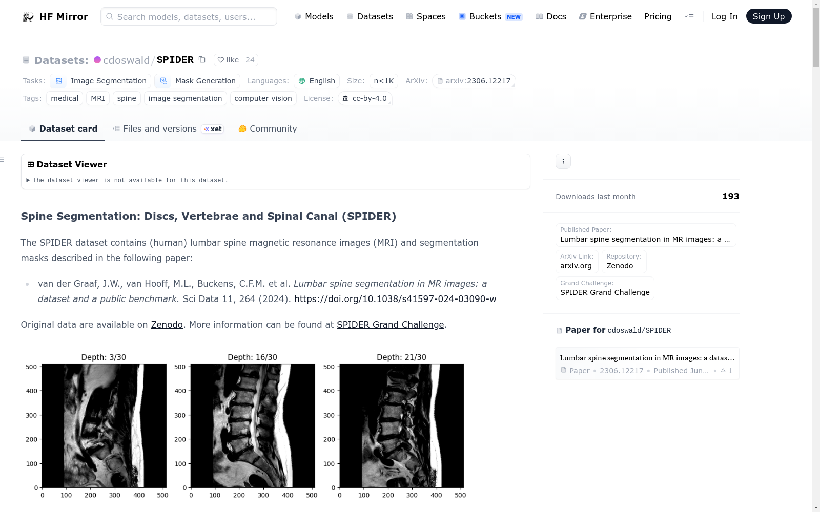
<figure>
<img src="docs/ex1.png" alt="Example MRI Image" style="height:300px;">
<figcaption>Example MRI scan (at three different depths)</figcaption>
</figure>
<figure>
<img src="docs/ex2.png" alt="Example MRI Image with Segmentation Mask" style="height:300px;">
<figcaption>Example MRI scan with segmentation masks</figcaption>
</figure>
# Dataset Description
- **Published Paper:** [Lumbar spine segmentation in MR images: a dataset and a public benchmark](https://www.nature.com/articles/s41597-024-03090-w)
- **ArXiv Link:** https://arxiv.org/abs/2306.12217
- **Repository:** [Zenodo](https://zenodo.org/records/8009680)
- **Grand Challenge:** [SPIDER Grand Challenge](https://spider.grand-challenge.org/)
# Tutorials
In addition to the information in this README, several detailed tutorials for this dataset are provided in the [tutorials](tutorials) folder:
1. [Loading the SPIDER Dataset from HuggingFace](tutorials/load_data.ipynb)
2. [Building a U-Net CNN Model for Magnetic Resonance Imaging (MRI) Segmentation](tutorials/UNet_SPIDER.ipynb)
<br>
# Table of Contents (TOC)
1. [Getting Started](https://huggingface.co/datasets/cdoswald/SPIDER#getting-started)
2. [Dataset Summary](https://huggingface.co/datasets/cdoswald/SPIDER#dataset-summary)
3. [Data Modifications](https://huggingface.co/datasets/cdoswald/SPIDER#data-modifications)
4. [Dataset Structure](https://huggingface.co/datasets/cdoswald/SPIDER#dataset-structure)
- [Data Instances](https://huggingface.co/datasets/cdoswald/SPIDER#data-instances)
- [Data Schema](https://huggingface.co/datasets/cdoswald/SPIDER#data-schema)
- [Data Splits](https://huggingface.co/datasets/cdoswald/SPIDER#data-splits)
5. [Image Resolution](https://huggingface.co/datasets/cdoswald/SPIDER#image-resolution)
6. [Additional Information](https://huggingface.co/datasets/cdoswald/SPIDER#additional-information)
- [License](https://huggingface.co/datasets/cdoswald/SPIDER#license)
- [Citation](https://huggingface.co/datasets/cdoswald/SPIDER#citation)
- [Disclaimer](https://huggingface.co/datasets/cdoswald/SPIDER#disclaimer)
- [Known Issues/Bugs](https://huggingface.co/datasets/cdoswald/SPIDER#known-issuesbugs)
<br>
# Getting Started
First, you will need to install the following dependencies:
* `datasets >= 2.18.0`
* `scikit-image >= 0.19.3`
* `SimpleITK >= 2.3.1`
Then you can load the SPIDER dataset as follows:
```python
from datasets import load_dataset
dataset = load_dataset("cdoswald/SPIDER, name="default", trust_remote_code=True)
```
See the [Loading the Dataset](tutorials/load_data.ipynb) tutorial for more information.
# Dataset Summary
The dataset includes 447 sagittal T1 and T2 MRI series collected from 218 patients across four hospitals.
Segmentation masks indicating the vertebrae, intervertebral discs (IVDs), and spinal canal are also included.
Segmentation masks were created manually by a medical trainee under the supervision of a medical imaging expert and an experienced musculoskeletal radiologist.
In addition to MR images and segmentation masks, additional metadata (e.g., scanner manufacturer, pixel bandwidth, etc.), limited
patient characteristics (biological sex and age, when available), and radiological gradings indicating specific degenerative
changes can be loaded with the corresponding image data.
# Data Modifications
This version of the SPIDER dataset (i.e., available through the HuggingFace `datasets` library) differs from the original
data available on [Zenodo](https://zenodo.org/records/8009680) in two key ways:
1. Image Rescaling/Resizing: The original 3D volumetric MRI data are stored as .mha files and do not have a standardized height, width, depth, and image resolution.
To enable the data to be loaded through the HuggingFace `datasets` library, all 447 MRI series are standardized to have height and width of `(512, 512)` and (unsigned) 16-bit integer resolution.
Segmentation masks have the same height and width dimension but are (unsigned) 8-bit integer resolution.
The depth dimension has not been modified; rather, each scan is formatted as a sequence of `(512, 512)` grayscale images, where the index in the sequence indicates the depth value.
N-dimensional interpolation is used to resize and/or rescale the images (via the `skimage.transform.resize` and `skimage.img_as_uint` functions).
If you need a different standardization, you have two options:
i. Pass your preferred height and width size as a `Tuple[int, int]` to the `resize_shape` argument in `load_dataset` (see the [LoadData Tutorial](placeholder)); OR
ii. After loading the dataset from HuggingFace, use the `SimpleITK` library to import each image using the file path of the locally cached .mha file.
The local cache file path is provided for each example when iterating over the dataset (again, see the [LoadData Tutorial](placeholder)).
2. Train, Validation, and Test Set: The original dataset contained 257 unique studies (i.e., patients) that were partitioned into 218 (85%) studies for the public training/validation set
and 39 (15%) studies for the SPIDER Grand Challenge [hidden test set](https://spider.grand-challenge.org/data/). To enable users to train, validate, and test their models prior to submitting
their models to the SPIDER Grand Challenge, the original 218 studies that comprised the public training/validation set were further partitioned using a 60%/20%/20% split. The original split
for each study (i.e., training or validation set) is recorded in the `OrigSubset` variable in the study's linked metadata.
# Dataset Structure
### Data Instances
There are 447 images and corresponding segmentation masks for 218 unique patients.
### Data Schema
The format for each generated data instance is as follows:
1. **patient_id**: a unique ID number indicating the specific patient (note that many patients have more than one scan in the data)
2. **scan_type**: an indicator for whether the image is a T1-weighted, T2-weighted, or T2-SPACE MRI
3. **image**: a sequence of 2-dimensional grayscale images of the MRI scan
4. **mask**: a sequence of 2-dimensional values indicating the following segmented anatomical feature(s):
- 0 = background
- 1-25 = vertebrae (numbered from the bottom, i.e., L5 = 1)
- 100 = spinal canal
- 101-125 = partially visible vertebrae
- 201-225 = intervertebral discs (numbered from the bottom, i.e., L5/S1 = 201)
See the [SPIDER Grand Challenge](https://grand-challenge.org/algorithms/spider-baseline-iis/) documentation for more details.
6. **image_path**: path to the local cache containing the original (non-rescaled and non-resized) MRI image
7. **mask_path**: path to the local cache containing the original (non-rescaled and non-resized) segementation mask
8. **metadata**: a dictionary of metadata of image, patient, and scanner characteristics:
- number of vertebrae
- number of discs
- biological sex
- age
- manufacturer
- manufacturer model name
- serial number
- software version
- echo numbers
- echo time
- echo train length
- flip angle
- imaged nucleus
- imaging frequency
- inplane phase encoding direction
- MR acquisition type
- magnetic field strength
- number of phase encoding steps
- percent phase field of view
- percent sampling
- photometric interpretation
- pixel bandwidth
- pixel spacing
- repetition time
- specific absorption rate (SAR)
- samples per pixel
- scanning sequence
- sequence name
- series description
- slice thickness
- spacing between slices
- specific character set
- transmit coil name
- window center
- window width
9. **rad_gradings**: radiological gradings by an expert musculoskeletal radiologist indicating specific degenerative
changes at all intervertebral disc (IVD) levels (see page 3 of the [original paper](https://www.nature.com/articles/s41597-024-03090-w)
for more details). The data are provided as a dictionary of lists; an element's position in the list indicates the IVD level. Some elements
are ratings while others are binary indicators. For consistency, each list will have 10 elements, but some IVD levels may not be applicable
to every image (which will be indicated with an empty string).
### Data Splits
The dataset is split as follows:
- Training set:
- 149 unique patients
- 304 total images
- Sagittal T1: 133 images
- Sagittal T2: 145 images
- Sagittal T2-SPACE: 26 images
- Validation set:
- 37 unique patients
- 75 total images
- Sagittal T1: 34 images
- Sagittal T2: 34 images
- Sagittal T2-SPACE: 7 images
- Test set:
- 32 unique patients
- 68 total images
- Sagittal T1: 29 images
- Sagittal T2: 31 images
- Sagittal T2-SPACE: 8 images
An additional hidden test set provided by the paper authors
(i.e., not available via HuggingFace) is available on the
[SPIDER Grand Challenge](https://spider.grand-challenge.org/spiders-challenge/).
# Image Resolution
> Standard sagittal T1 and T2 image resolution ranges from 3.3 x 0.33 x 0.33 mm to 4.8 x 0.90 x 0.90 mm.
> Sagittal T2 SPACE sequence images had a near isotropic spatial resolution with a voxel size of 0.90 x 0.47 x 0.47 mm.
> (https://spider.grand-challenge.org/data/)
Note that all images are rescaled to have unsigned 16-bit integer resolution
for compatibility with the HuggingFace `datasets` library. If you want to use the original resolution, you can
load the original images from the local cache indicated in each example's `image_path` and `mask_path` features.
See the [tutorial](tutorials/load_data.ipynb) for more information.
# Additional Information
### License
The dataset is published under a CC-BY 4.0 license: https://creativecommons.org/licenses/by/4.0/legalcode.
### Citation
- van der Graaf, J.W., van Hooff, M.L., Buckens, C.F.M. et al. Lumbar spine segmentation in MR images: a dataset and a public benchmark. Sci Data 11, 264 (2024). https://doi.org/10.1038/s41597-024-03090-w.
### Disclaimer
I am not affiliated in any way with the aforementioned paper, researchers, or organizations. Please validate any findings using this curated dataset
against the original data provided by the researchers on [Zenodo](https://zenodo.org/records/10159290).
### Known Issues/Bugs
1. Serializing data into Apache Arrow format is required to make the dataset available via HuggingFace's `datasets` library. However, it can introduce some segmentation
mask integer values that do not map exactly to a defined [anatomical feature category](https://grand-challenge.org/algorithms/spider-baseline-iis/).
See the data loading [tutorial](tutorials/load_data.ipynb) for more information and temporary work-arounds.
---
许可证:CC-BY-4.0
语言:
- 英语
标签:
- 医学
- 磁共振成像(MRI)
- 脊柱
- 图像分割
- 计算机视觉
样本规模:
- 少于1000
美观名称:SPIDER:脊柱MRI分割
任务类别:
- 图像分割
- 掩码生成
---
# 脊柱分割:椎间盘、椎体与椎管(SPIDER)
SPIDER数据集包含人类腰椎磁共振成像(MRI)与分割掩码,相关细节详见以下论文:
- van der Graaf, J.W., van Hooff, M.L., Buckens, C.F.M. 等人:《MR图像中的腰椎分割:数据集与公开基准》,《科学数据》(Sci Data)11卷,第264页(2024年),DOI: 10.1038/s41597-024-03090-w
原始数据可在[Zenodo](https://zenodo.org/records/10159290)获取,更多信息可查阅[SPIDER大赛官网](https://spider.grand-challenge.org/)。
<figure>
<img src="docs/ex1.png" alt="示例MRI图像" style="height:300px;">
<figcaption>示例MRI扫描图像(三种不同层面)</figcaption>
</figure>
<figure>
<img src="docs/ex2.png" alt="带分割掩码的MRI图像示例" style="height:300px;">
<figcaption>带分割掩码的MRI扫描示例图像</figcaption>
</figure>
# 数据集说明
- **已发表论文**:[《MR图像中的腰椎分割:数据集与公开基准》](https://www.nature.com/articles/s41597-024-03090-w)
- **ArXiv链接**:https://arxiv.org/abs/2306.12217
- **代码仓库**:[Zenodo](https://zenodo.org/records/8009680)
- **大赛官网**:[SPIDER大赛官网](https://spider.grand-challenge.org/)
# 教程
除本README文档外,本数据集的多份详细教程已存放于[tutorials](tutorials)文件夹中:
1. [从HuggingFace加载SPIDER数据集](tutorials/load_data.ipynb)
2. [构建用于磁共振成像(MRI)分割的U-Net卷积神经网络模型](tutorials/UNet_SPIDER.ipynb)
<br>
# 目录(TOC)
1. [快速入门](https://huggingface.co/datasets/cdoswald/SPIDER#getting-started)
2. [数据集概述](https://huggingface.co/datasets/cdoswald/SPIDER#dataset-summary)
3. [数据修改说明](https://huggingface.co/datasets/cdoswald/SPIDER#data-modifications)
4. [数据集结构](https://huggingface.co/datasets/cdoswald/SPIDER#dataset-structure)
- [数据实例](https://huggingface.co/datasets/cdoswald/SPIDER#data-instances)
- [数据格式](https://huggingface.co/datasets/cdoswald/SPIDER#data-schema)
- [数据划分](https://huggingface.co/datasets/cdoswald/SPIDER#data-splits)
5. [图像分辨率](https://huggingface.co/datasets/cdoswald/SPIDER#image-resolution)
6. [附加信息](https://huggingface.co/datasets/cdoswald/SPIDER#additional-information)
- [许可证](https://huggingface.co/datasets/cdoswald/SPIDER#license)
- [引用格式](https://huggingface.co/datasets/cdoswald/SPIDER#citation)
- [免责声明](https://huggingface.co/datasets/cdoswald/SPIDER#disclaimer)
- [已知问题与缺陷](https://huggingface.co/datasets/cdoswald/SPIDER#known-issuesbugs)
<br>
# 快速入门
首先,请安装以下依赖项:
* `datasets >= 2.18.0`
* `scikit-image >= 0.19.3`
* `SimpleITK >= 2.3.1`
随后即可通过如下方式加载SPIDER数据集:
python
from datasets import load_dataset
dataset = load_dataset("cdoswald/SPIDER", name="default", trust_remote_code=True)
更多细节请参阅[数据集加载教程](tutorials/load_data.ipynb)。
# 数据集概述
本数据集包含来自4家医院的218名患者的447组矢状位T1、T2加权MRI序列。同时包含标注椎体、椎间盘(IVDs)与椎管的分割掩码。
分割掩码由医学规培生在医学影像专家与资深肌肉骨骼放射科医师的监督下手动标注完成。
除MRI图像与分割掩码外,配套加载的还包括各类元数据(如扫描仪厂商、像素带宽等)、有限的患者特征信息(如可获取的生物性别与年龄),以及指示特定退行性改变的放射学分级数据。
# 数据修改说明
本版本的SPIDER数据集(即可通过HuggingFace `datasets`库获取的版本)与[Zenodo](https://zenodo.org/records/8009680)上的原始数据存在两处关键差异:
1. 图像重缩放与尺寸调整:原始3D体素MRI数据以.mha格式存储,未统一高度、宽度、深度与图像分辨率。为适配HuggingFace `datasets`库的加载要求,本版本将全部447组MRI序列的高与宽统一调整为`(512, 512)`,并转换为无符号16位整数精度。分割掩码保持相同的高宽尺寸,但采用无符号8位整数精度。深度维度未做修改,每组扫描均格式化为`(512, 512)`灰度图像序列,序列索引对应层面深度。图像的尺寸调整与重缩放采用N维插值实现(通过`skimage.transform.resize`与`skimage.img_as_uint`函数完成)。若需采用其他标准化方案,可通过两种方式实现:
i. 在调用`load_dataset`时,将自定义的高宽尺寸以`Tuple[int, int]`格式传入`resize_shape`参数(详见[数据加载教程](placeholder));或
ii. 从HuggingFace加载数据集后,通过`SimpleITK`库,利用本地缓存的.mha文件路径导入原始图像。遍历数据集时,每个样本均会提供本地缓存文件路径(再次详见[数据加载教程](placeholder))。
2. 训练集、验证集与测试集划分:原始数据集包含257个独立研究(即患者),原划分为218个(85%)研究作为公开训练/验证集,39个(15%)研究作为SPIDER大赛的[隐藏测试集](https://spider.grand-challenge.org/data/)。为方便用户在提交模型至SPIDER大赛前自行开展训练、验证与测试,原公开训练/验证集的218个研究被进一步按60%/20%/20%的比例划分为训练、验证与测试集。每个研究的原始划分(训练集或验证集)记录在对应元数据的`OrigSubset`变量中。
# 数据集结构
### 数据实例
本数据集包含218名独立患者的447组图像及其对应分割掩码。
### 数据格式
每个生成的数据样本格式如下:
1. **patient_id**:用于标识特定患者的唯一ID编号(请注意,多位患者在数据中包含多组扫描序列)
2. **scan_type**:标识图像类型,包括T1加权、T2加权或T2-SPACE MRI
3. **image**:MRI扫描的二维灰度图像序列
4. **mask**:二维数值序列,用于标注以下分割解剖结构:
- 0 = 背景
- 1-25 = 椎体(从下往上编号,即L5 = 1)
- 100 = 椎管
- 101-125 = 部分可见椎体
- 201-225 = 椎间盘(从下往上编号,即L5/S1 = 201)
更多细节请参阅[SPIDER大赛官网](https://grand-challenge.org/algorithms/spider-baseline-iis/)的文档说明。
6. **image_path**:指向本地缓存的路径,存储未经过重缩放与尺寸调整的原始MRI图像
7. **mask_path**:指向本地缓存的路径,存储未经过重缩放与尺寸调整的原始分割掩码
8. **metadata**:包含图像、患者与扫描仪特征的元数据字典:
- 椎体数量
- 椎间盘数量
- 生物性别
- 年龄
- 扫描仪厂商
- 扫描仪型号
- 序列号
- 软件版本
- 回波数
- 回波时间
- 回波链长度
- 翻转角
- 成像核
- 成像频率
- 平面内相位编码方向
- MRI采集类型
- 磁场强度
- 相位编码步数
- 相位视场百分比
- 采样百分比
- 光度解释
- 像素带宽
- 像素间距
- 重复时间
- 比吸收率(SAR)
- 每像素采样数
- 扫描序列
- 序列名称
- 序列描述
- 层厚
- 层间距
- 特定字符集
- 发射线圈名称
- 窗位
- 窗宽
9. **rad_gradings**:由资深肌肉骨骼放射科医师标注的放射学分级数据,用于指示所有椎间盘(IVD)节段的特定退行性改变(更多细节请参阅[原论文](https://www.nature.com/articles/s41597-024-03090-w)第3页)。该数据以字典嵌套列表的形式提供,列表中元素的位置对应椎间盘节段。部分元素为分级评分,部分为二元指示符。为保持格式统一,每个列表均包含10个元素,但部分椎间盘节段可能不适用于某组图像,此时将以空字符串标注。
### 数据划分
本数据集划分如下:
- 训练集:
- 149名独立患者
- 共304组图像
- 矢状位T1加权:133组
- 矢状位T2加权:145组
- 矢状位T2-SPACE:26组
- 验证集:
- 37名独立患者
- 共75组图像
- 矢状位T1加权:34组
- 矢状位T2加权:34组
- 矢状位T2-SPACE:7组
- 测试集:
- 32名独立患者
- 共68组图像
- 矢状位T1加权:29组
- 矢状位T2加权:31组
- 矢状位T2-SPACE:8组
论文作者还提供了额外的隐藏测试集(即无法通过HuggingFace获取),可在[SPIDER大赛官网](https://spider.grand-challenge.org/spiders-challenge/)获取。
# 图像分辨率
> 标准矢状位T1与T2加权图像的分辨率范围为3.3×0.33×0.33 mm至4.8×0.90×0.90 mm。
> 矢状位T2 SPACE序列图像的空间分辨率接近各向同性,体素尺寸为0.90×0.47×0.47 mm。
> (来源:https://spider.grand-challenge.org/data/)
请注意,为适配HuggingFace `datasets`库,本版本所有图像均被重缩放为无符号16位整数精度。若需使用原始分辨率,可通过每个样本的`image_path`与`mask_path`字段指向的本地缓存加载原始图像。更多细节请参阅[教程](tutorials/load_data.ipynb)。
# 附加信息
### 许可证
本数据集采用CC-BY 4.0许可证发布:https://creativecommons.org/licenses/by/4.0/legalcode。
### 引用格式
- van der Graaf, J.W.、van Hooff, M.L.、Buckens, C.F.M. 等人:《MR图像中的腰椎分割:数据集与公开基准》,《科学数据》11卷,第264页(2024年),DOI: 10.1038/s41597-024-03090-w。
### 免责声明
本人与上述论文、研究人员及相关组织无任何关联。若使用本整理后的数据集获得研究结果,请务必与研究者在[Zenodo](https://zenodo.org/records/10159290)发布的原始数据进行比对验证。
### 已知问题与缺陷
1. 为使数据集可通过HuggingFace的`datasets`库获取,需将数据序列化为Apache Arrow格式,但该过程可能导致部分分割掩码的整数值无法与已定义的[解剖结构类别](https://grand-challenge.org/algorithms/spider-baseline-iis/)完全匹配。更多细节与临时解决方案请参阅数据加载[教程](tutorials/load_data.ipynb)。
提供机构:
cdoswald
原始信息汇总
SPIDER: Spine MRI Segmentation 数据集概述
数据集基本信息
- 许可证: CC-BY 4.0
- 语言: 英语
- 标签: 医学, MRI, 脊柱, 图像分割, 计算机视觉
- 数据集大小: n<1K
- 数据集名称: SPIDER: Spine MRI Segmentation
- 任务类别: 图像分割, 掩码生成
数据集描述
SPIDER 数据集包含人类腰椎磁共振图像(MRI)及其分割掩码,详细描述见以下论文:
- van der Graaf, J.W., van Hooff, M.L., Buckens, C.F.M. et al. Lumbar spine segmentation in MR images: a dataset and a public benchmark. Sci Data 11, 264 (2024). https://doi.org/10.1038/s41597-024-03090-w
数据集内容
- 论文链接: Lumbar spine segmentation in MR images: a dataset and a public benchmark
- ArXiv 链接: https://arxiv.org/abs/2306.12217
- 数据存储库: Zenodo
- 挑战赛链接: SPIDER Grand Challenge
数据集结构
- 数据实例: 包含 447 张图像和相应的分割掩码,对应 218 名独特的患者。
- 数据模式:
- patient_id: 患者唯一 ID 号
- scan_type: 图像类型(T1-weighted, T2-weighted, T2-SPACE MRI)
- image: MRI 扫描的二维灰度图像序列
- mask: 分割掩码,指示以下解剖特征:
- 0 = 背景
- 1-25 = 椎骨(从底部编号,L5 = 1)
- 100 = 脊髓管
- 101-125 = 部分可见的椎骨
- 201-225 = 椎间盘(从底部编号,L5/S1 = 201)
- image_path: 原始(未重新缩放和调整大小)MRI 图像的本地缓存路径
- mask_path: 原始(未重新缩放和调整大小)分割掩码的本地缓存路径
- metadata: 图像、患者和扫描仪特征的元数据字典
- rad_gradings: 专家肌肉骨骼放射科医生对所有椎间盘水平的特定退行性变化的放射学评分
数据分割
- 训练集: 149 名独特患者,304 张图像
- 验证集: 37 名独特患者,75 张图像
- 测试集: 32 名独特患者,68 张图像
- 隐藏测试集: 由论文作者提供的额外隐藏测试集,不可通过 HuggingFace 获取
图像分辨率
- 标准矢状 T1 和 T2 图像分辨率范围从 3.3 x 0.33 x 0.33 mm 到 4.8 x 0.90 x 0.90 mm。
- 矢状 T2 SPACE 序列图像具有近各向同性的空间分辨率,体素大小为 0.90 x 0.47 x 0.47 mm。
数据集修改
- 图像重新缩放/调整大小: 所有 447 个 MRI 系列标准化为高度和宽度
(512, 512),分割掩码具有相同的高度和宽度维度,但为无符号 8 位整数分辨率。 - 训练、验证和测试集: 原始数据集被进一步分割为 60%/20%/20% 的比例。
许可证
- 数据集发布在 CC-BY 4.0 许可证下:https://creativecommons.org/licenses/by/4.0/legalcode
引用
- van der Graaf, J.W., van Hooff, M.L., Buckens, C.F.M. et al. Lumbar spine segmentation in MR images: a dataset and a public benchmark. Sci Data 11, 264 (2024). https://doi.org/10.1038/s41597-024-03090-w.
免责声明
- 与上述论文、研究人员或组织无任何关联。请在使用此数据集时验证任何发现与研究人员在 Zenodo 上提供的原始数据。
已知问题/错误
- 序列化数据到 Apache Arrow 格式时,可能会引入一些分割掩码整数值,这些值不完全映射到定义的解剖特征类别。
搜集汇总
数据集介绍
背景与挑战
背景概述
SPIDER数据集是一个专注于腰椎MRI图像分割的医学影像数据集,包含447个来自218名患者的MRI图像和手动标注的分割掩码,适用于计算机视觉和医学图像分析任务。数据集经过标准化处理,提供了详细的元数据和放射学评分,支持训练、验证和测试集的划分,旨在促进腰椎分割算法的研究和开发。
以上内容由遇见数据集搜集并总结生成


