基于多模式MRI的狨猴大脑数字三维图谱
收藏中国科学院脑科学数据中心2022-10-27 更新2024-03-05 收录
下载链接:
https://www.braindatacenter.cn/datacenter/web/#/dataSet/details?id=1585637188254449665
下载链接
链接失效反馈官方服务:
资源简介:
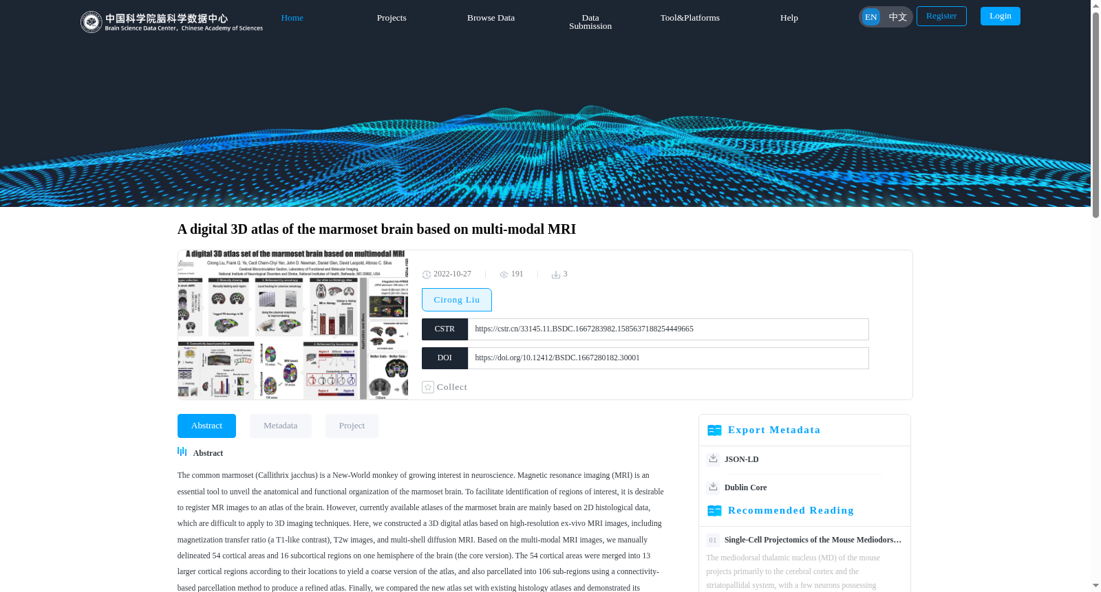
普通狨猴(Callithrix jacchus)是一种新兴的模式动物,在神经科学方面的兴趣越来越大。磁共振成像(MRI)是揭示狨猴大脑的解剖和功能组织的一个重要工具。为了便于识别感兴趣的区域,最好将磁共振图像登记到大脑的图谱上。然而,目前可用的狨猴大脑图谱主要基于二维组织学数据,难以应用于三维成像技术。在这里,我们根据高分辨率的体外MRI图像,包括磁化转移率(一种类似T1的对比)、T2w图像和多壳扩散MRI,构建了一个三维数字图谱。基于多模式的MRI图像,我们手动划定了大脑一个半球上的54个皮质区域和16个皮质下区域(核心版)。54个皮层区域根据其位置被合并成13个较大的皮层区域,以产生一个粗略版本的图谱,并使用基于连接的分割方法将其分割成106个亚区域,以产生一个精细的图谱。最后,我们将新的图集与现有的组织学图集进行了比较,并展示了其在连接组研究、静止状态和基于刺激的fMRI中的应用。该图集已被整合到广泛分布的神经影像数据分析软件AFNI和SUMA中,提供了一个具有多级解剖学标签(包括来自Paxinos图集的标签)的随时可用的多模式模板空间,可以促进狨猴的各种神经影像学研究。
The common marmoset (*Callithrix jacchus*) has emerged as an increasingly popular model organism in neuroscience research. Magnetic resonance imaging (MRI) is a critical tool for revealing the anatomical and functional organization of the marmoset brain. To facilitate the identification of regions of interest (ROIs), it is optimal to register MRI images to a brain atlas. However, currently available marmoset brain atlases are primarily based on 2D histological data, making them difficult to apply to 3D imaging technologies. Here, we constructed a 3D digital atlas based on high-resolution ex vivo MRI images, including magnetization transfer ratio (MTR, a T1-like contrast), T2-weighted (T2w) images, and multi-shell diffusion MRI. Based on these multimodal MRI images, we manually annotated 54 cortical regions and 16 subcortical regions (core version) on one hemisphere of the marmoset brain. The 54 cortical regions were merged into 13 larger cortical regions based on their locations to generate a coarse atlas, and were also segmented into 106 subregions using a connection-based segmentation method to produce a fine-grained atlas. Finally, we compared the new atlas with existing histological atlases, and demonstrated its applications in connectomics research, resting-state fMRI, and stimulus-evoked fMRI studies. This atlas has been integrated into widely used neuroimaging data analysis software AFNI and SUMA, providing a ready-to-use multimodal template space with multi-level anatomical labels (including labels from the Paxinos atlas), which can facilitate various neuroimaging studies on marmosets.
提供机构:
中国科学院脑科学数据中心
创建时间:
2022-10-27
搜集汇总
数据集介绍
背景与挑战
背景概述
该数据集是一个基于多模式MRI构建的狨猴大脑数字3D图谱,包含高分辨率离体MRI图像和手动描绘的脑区标签,提供核心、粗粒度和细化三个版本。它旨在解决现有2D组织学图谱难以应用于3D成像技术的问题,已集成到AFNI和SUMA等神经影像分析软件中,支持狨猴大脑的连接组学、功能MRI等研究。
以上内容由遇见数据集搜集并总结生成


