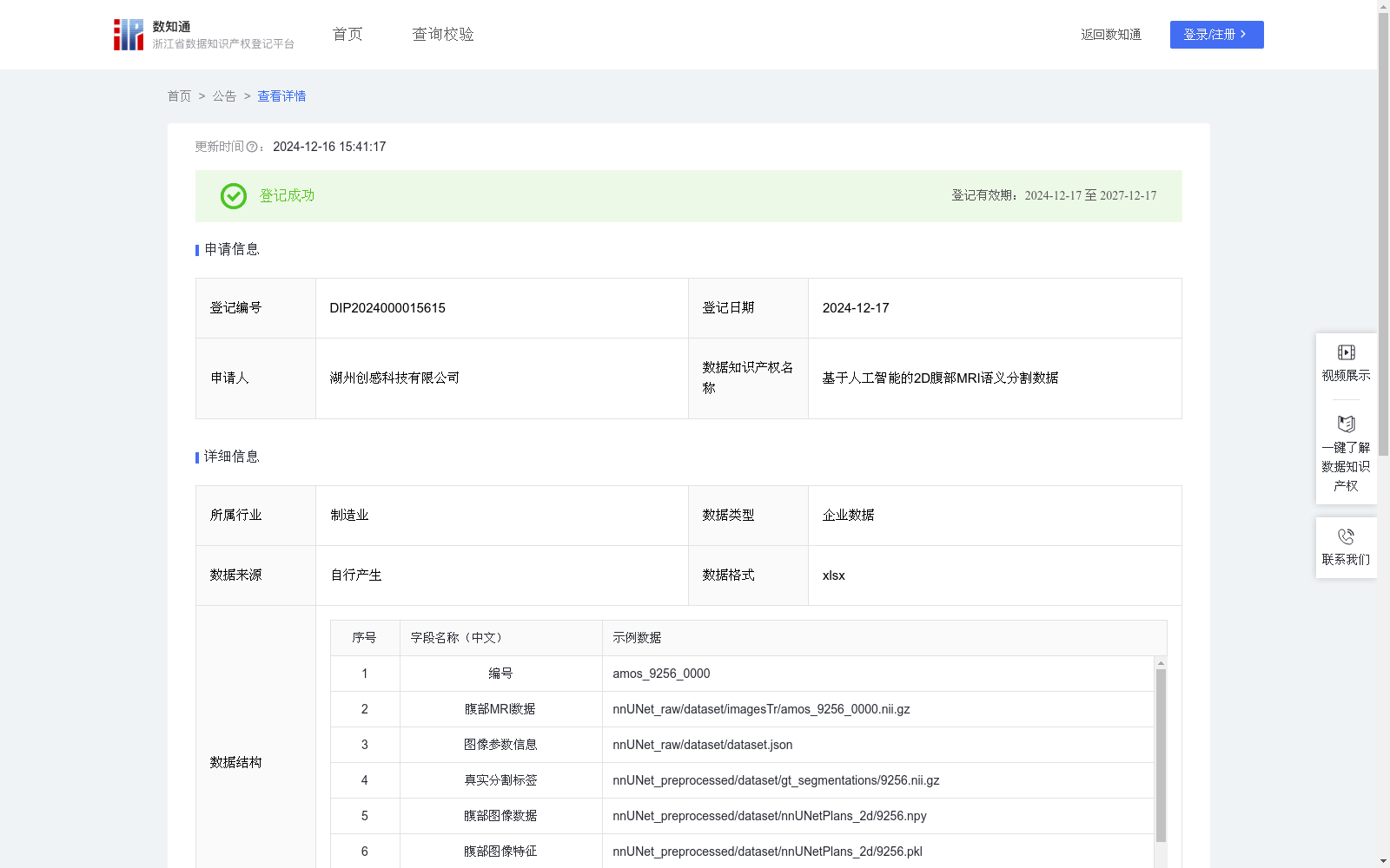
基于人工智能的2D腹部MRI语义分割数据
收藏浙江省数据知识产权登记平台2024-12-16 更新2024-12-17 收录
下载链接:
https://www.zjip.org.cn/home/announce/trends/104917
下载链接
链接失效反馈官方服务:
资源简介:
基于人工智能的二维腹部MRI语义分割技术广泛应用于医学影像分析领域,尤其是在临床诊断、疾病检测、治疗规划等方面。在腹部MRI图像中,精确地分割不同的组织结构,如肝脏、肾脏、肠道等,对于医生来说是一个挑战。通过结合深度学习模型进行语义分割,可以自动化地提取腹部MRI图像中的关键区域,提高诊断的效率和精度。这项技术对于临床医学、放射学、病理学等专业领域尤其重要,能够帮助医生更快地识别病变部位,支持个性化治疗方案的制定。在医学研究领域,它可以辅助分析疾病的进展和预测治疗效果,推动医学影像智能化的发展。数据收集:在该算法中,首先收集腹部MRI数据以及相应的真实分割标签,作为模型训练和验证的基础。每个病例包括:腹部MRI数据(*.nii.gz格式文件),包含腹部MRI的原始图像数据。图像参数信息(*.json格式),用于记录MRI图像的具体参数,帮助模型更好地理解图像的特征。真实分割标签,表示不同组织或病变区域的真实标注信息,作为监督学习的目标数据。
数据预处理:在数据预处理阶段,腹部MRI数据(.nii.gz格式)首先需要处理为腹部图像数据,并进行标准化处理,包括缩放、归一化等步骤,以适应神经网络的输入要求。接着,将处理后的腹部图像数据转化为适用于训练的格式。处理过程还包括数据增强,如旋转、翻转、裁剪等,帮助模型学习更多变形的腹部图像特征,提高其泛化能力。
模型构建:使用深度学习网络进行图像的语义分割。网络输入为预处理后的图像数据,输出为对不同器官的分割标签。具体算法公式如下:F_features = Encoder_features(Processed_image),Output_segmentation = Decoder_segmentation(F_features)。其中,Encoder_features为图像特征提取网络,Decoder_segmentation是用于生成分割标签的解码器。网络通过训练学习腹部MRI图像的关键特征,并生成对应的预测分割标签。最后使用DSC和NSD指标进行评估。
AI-based 2D abdominal MRI semantic segmentation technology is widely used in medical image analysis, especially in clinical diagnosis, disease detection, treatment planning and other fields.
Accurately segmenting different anatomical structures such as the liver, kidney, intestine and other tissues in abdominal MRI images poses a challenge for clinicians.
Combining deep learning models for semantic segmentation enables automated extraction of key regions from abdominal MRI images, improving the efficiency and accuracy of diagnosis.
This technology is particularly important in professional fields such as clinical medicine, radiology and pathology, as it helps clinicians identify lesion sites faster and supports the development of personalized treatment plans.
In medical research, it can assist in analyzing disease progression and predicting treatment outcomes, promoting the development of intelligent medical imaging.
Data Collection: In this algorithm, abdominal MRI data and corresponding ground-truth segmentation labels are first collected as the foundation for model training and validation.
Each case includes:
1. Abdominal MRI data (files in *.nii.gz format), which contains the original abdominal MRI image data.
2. Image parameter information (in *.json format), used to record specific parameters of the MRI image to help the model better understand image features.
3. Ground-truth segmentation labels, which represent real annotation information of different tissues or lesion regions, serving as the target data for supervised learning.
Data Preprocessing: In the data preprocessing stage, abdominal MRI data (in *.nii.gz format) must first be processed into standard abdominal image data and undergo standardization operations including scaling, normalization and other steps to meet the input requirements of neural networks.
Subsequently, the processed abdominal image data is converted into a format suitable for model training.
The preprocessing process also includes data augmentation techniques such as rotation, flipping, cropping and others, which help the model learn more varied abdominal image features and improve its generalization ability.
Model Construction: A deep learning network is used for image semantic segmentation.
The network takes the preprocessed image data as input and outputs segmentation labels for different organs.
The specific algorithm formulas are as follows:
$$F_{features} = Encoder_features(Processed_image)$$
$$Output_{segmentation} = Decoder_segmentation(F_{features})$$
Here, $Encoder_features$ refers to the image feature extraction network, and $Decoder_segmentation$ is the decoder used to generate segmentation labels.
The network learns the key features of abdominal MRI images through training and generates corresponding predicted segmentation labels.
Finally, the Dice Similarity Coefficient (DSC) and Normalized Surface Dice (NSD) metrics are used for model evaluation.
提供机构:
湖州创感科技有限公司
创建时间:
2024-11-14
搜集汇总
数据集介绍
以上内容由遇见数据集搜集并总结生成


