盆底超声影像AI诊断分析数据
收藏浙江省数据知识产权登记平台2025-09-30 更新2025-10-04 收录
下载链接:
https://www.zjip.org.cn/home/announce/trends/187951
下载链接
链接失效反馈官方服务:
资源简介:
一、适用条件与对象
适用于各级医院妇产科及超声科,特别针对产后42天至1年内的产妇及老年女性群体的盆底功能筛查。服务对象包括临床医师、康复治疗师及医疗AI研发机构。
二、解决的问题和痛点
(1)解决盆底超声诊断高度依赖医师经验、结构识别一致性差的问题;
(2)突破传统测量方法效率低下、量化指标提取困难的瓶颈;
(3)缓解基层医院盆底诊疗能力不足的困境。
三、有益效果
(1)为临床提供精准的盆底结构自动识别与量化评估(如肛提肌裂孔面积、膀胱颈移动度),生成客观的AI辅助诊断建议;
(2)本数据可提升诊断效率,显著降低漏诊率。
四、外部复用价值
(1)核心算法可集成至超声设备及PACS系统,实现"设备+AI"一体化解决方案;
(2)脱敏盆底特征数据库支持多中心科研与算法迭代;
(3)标准化的盆底超声影像AI诊断体系可推广至医联体,促进盆底诊疗规范化与优质资源下沉。一、数据采集
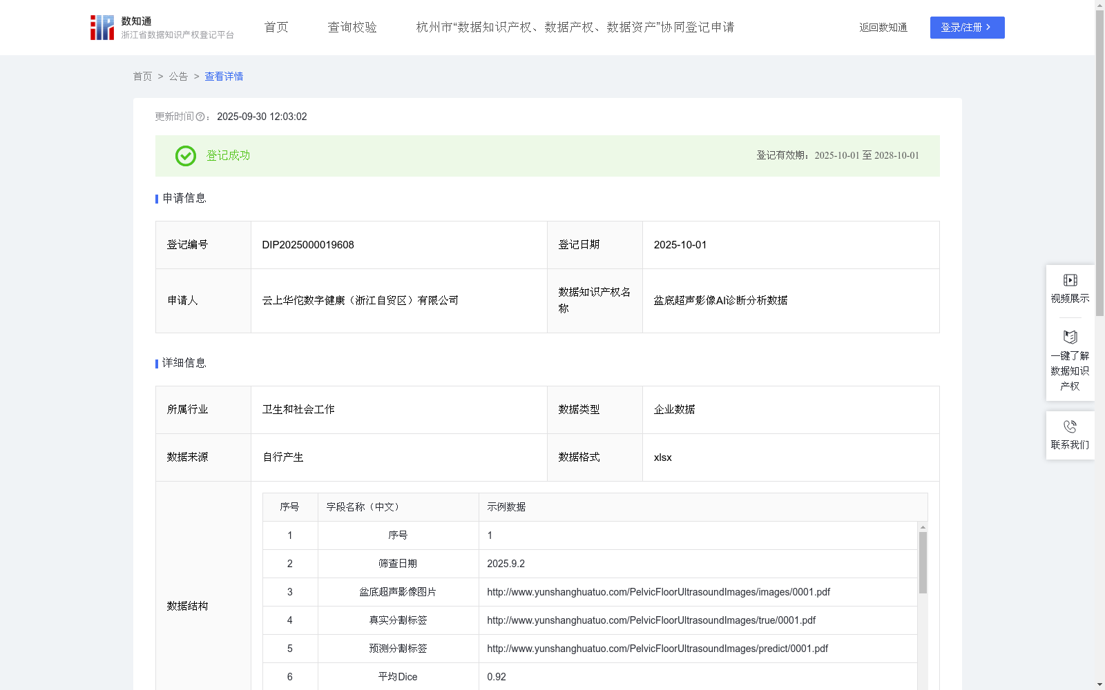
超声探头发射的超声波穿透盆底组织,遇到不同密度的组织界面时反射信号,这些信号被探头接收后转化为电信号,最终生成盆底超声影像图片,清晰呈现盆底肌肉、韧带、子宫、膀胱等结构的形态和位置,收集盆底超声影像相关的结构化特征信息。
核心字段:
盆底超声影像图片:通过URL存储盆底超声影像图片(URL链接已脱敏)
真实分割标签:专家标注的真实分割标签(精确诊断女性盆底功能障碍性疾病)
预测分割标签:模型识别的预测分割标签
盆底特征数据:子宫大小、宫内膜厚度、左卵巢直径、右卵巢直径、膀胱颈移动度、肛提肌裂孔面积、子宫形态与大小、盆底结构(前腔室)、盆底结构(中腔室)、盆底结构(后腔室)
诊断数据:AI辅助诊断建议
辅助字段:
临床建议、随访建议用于人工复核。筛查日期用于数据管理。
二、数据处理
对所有盆底超声影像图片进行标准化预处理:尺寸归一化(统一超声影像图片分辨率)、图像去噪(减少超声影像斑点噪声)、亮度对比度调整(优化超声影像质量)、特征数据向量化处理
目标:提升后续算法模型的鲁棒性和分割精度
三、核心算法规则 (模型构建与训练)
(1)模型架构:
采用编码器-解码器结构的卷积神经网络(CNN)
模型公式:P=CNNθ(I)
I:预处理后的盆底超声影像图片
CNNθ:卷积神经网络模型参数
P:女性盆底功能障碍性疾病的预测分割标签图
(2)训练策略:
损失函数:结合Dice损失和二元交叉熵损失
优化器:Adam
性能评估指标:平均Dice系数、平均IoU。在训练和验证阶段,使用平均Dice系数和平均IoU作为核心指标,量化模型预测分割标签结果与真实分割标签之间的重叠精度。
(3)多特征融合:
将盆底超声影像特征与临床盆底结构特征(膀胱、卵巢、子宫、肛提肌)融合,输出AI辅助诊断建议。
四、数据应用
(1)临床诊断辅助:自动生成女性盆底功能障碍性疾病分割结果及量化参数,输出AI辅助诊断建议;
(2)质量控制:实时监测分割质量(平均Dice≥0.80,平均IoU≥0.70),异常案例自动标记复核;
(3)科研价值:脱敏特征数据库支持模型迭代优化,多中心研究数据标准化支持。
1. Application Scope and Target Groups
Applicable to obstetrics and gynecology departments and ultrasound departments of hospitals at all levels, especially for pelvic floor function screening of puerperae from 42 days to 1 year postpartum and elderly female populations. The service targets include clinicians, rehabilitation therapists, and medical AI R&D institutions.
2. Solved Problems and Pain Points
(1) Addresses the issue that pelvic floor ultrasound diagnosis highly relies on physician experience, leading to poor consistency in structural recognition;
(2) Breaks through the bottlenecks of low efficiency of traditional measurement methods and difficulty in extracting quantitative indicators;
(3) Alleviates the dilemma of insufficient pelvic floor diagnosis and treatment capabilities in primary hospitals.
3. Beneficial Effects
(1) Provides clinicians with accurate automatic recognition and quantitative assessment of pelvic floor structures (e.g., levator hiatus area, bladder neck mobility), and generates objective AI-assisted diagnostic suggestions;
(2) This dataset can improve diagnostic efficiency and significantly reduce the missed diagnosis rate.
4. External Reusability Value
(1) The core algorithm can be integrated into ultrasound equipment and Picture Archiving and Communication Systems (PACS) to realize an "equipment + AI" integrated solution;
(2) The de-identified pelvic floor feature database supports multi-center scientific research and algorithm iteration;
(3) The standardized pelvic floor ultrasound imaging AI diagnosis system can be promoted to medical consortia, promoting the standardization of pelvic floor diagnosis and treatment and the downward transfer of high-quality medical resources.
5. Data Collection
Ultrasound waves emitted by the ultrasound probe penetrate pelvic floor tissues, and reflect signals when encountering tissue interfaces with different densities. These signals are received by the probe, converted into electrical signals, and finally generate pelvic floor ultrasound images, which clearly show the morphology and position of structures such as pelvic floor muscles, ligaments, uterus, and bladder. Structured feature information related to pelvic floor ultrasound imaging is collected.
Core Fields:
- Pelvic floor ultrasound images: Stored via URLs (URL links have been de-identified)
- Ground truth segmentation labels: Real segmentation labels annotated by experts (for accurate diagnosis of female pelvic floor dysfunction)
- Predicted segmentation labels: Predicted segmentation labels identified by the model
- Pelvic floor feature data: Uterus size, endometrial thickness, left ovarian diameter, right ovarian diameter, bladder neck mobility, levator hiatus area, uterine morphology and size, pelvic floor structures (anterior compartment), pelvic floor structures (middle compartment), pelvic floor structures (posterior compartment)
- Diagnostic data: AI-assisted diagnostic suggestions
Auxiliary Fields:
Clinical recommendations and follow-up recommendations for manual review; Screening date for data management.
6. Data Processing
Standardized preprocessing is performed on all pelvic floor ultrasound images: size normalization (unifying the resolution of ultrasound images), image denoising (reducing speckle noise in ultrasound images), brightness and contrast adjustment (optimizing ultrasound image quality), and vectorization processing of feature data.
Goal: Improve the robustness and segmentation accuracy of subsequent algorithm models.
7. Core Algorithm Rules (Model Construction and Training)
(1) Model Architecture:
An encoder-decoder structured convolutional neural network (CNN) is adopted.
Model formula: $P = CNN_ heta(I)$
Where:
$I$: Preprocessed pelvic floor ultrasound images
$CNN_ heta$: Convolutional neural network model parameters
$P$: Predicted segmentation label map for female pelvic floor dysfunction
(2) Training Strategy:
Loss function: Combines Dice loss and binary cross-entropy loss
Optimizer: Adam
Performance evaluation metrics: Average Dice coefficient, average Intersection over Union (IoU). In the training and validation stages, average Dice coefficient and average IoU are used as core metrics to quantify the overlap accuracy between the model's predicted segmentation label results and the ground truth segmentation labels.
(3) Multi-feature Fusion:
Fuse pelvic floor ultrasound imaging features with clinical pelvic floor structural features (bladder, ovary, uterus, levator ani muscle) to output AI-assisted diagnostic suggestions.
8. Data Application
(1) Clinical Diagnosis Assistance: Automatically generate segmentation results and quantitative parameters of female pelvic floor dysfunction, and output AI-assisted diagnostic suggestions;
(2) Quality Control: Real-time monitor segmentation quality (average Dice ≥ 0.80, average IoU ≥ 0.70), and automatically mark abnormal cases for review;
(3) Scientific Research Value: The de-identified feature database supports iterative optimization of the model, and provides support for standardized multi-center research data.
提供机构:
云上华佗数字健康(浙江自贸区)有限公司
创建时间:
2025-09-05
搜集汇总
数据集介绍
背景与挑战
背景概述
该数据集包含1923条盆底超声影像AI诊断分析数据,每日更新,涵盖盆底结构特征、分割标签和AI辅助诊断建议,用于训练卷积神经网络模型,自动识别女性盆底功能障碍性疾病。数据集通过量化指标(如平均Dice和IoU)评估模型性能,旨在解决临床诊断依赖经验、效率低下的痛点,提升诊断准确性和基层医疗能力。
以上内容由遇见数据集搜集并总结生成


