
Nakamura, K., Okamoto, M., Mada, T., Harada, M., Okumura, K., & Takamatsu, D. Data sets for comparison of the virulence of <i>Paenibacillus</i> sp. J27TS7 and <i>Paenibacillus melissococcoides</i> strains to honeybee larvae (Bioassay III).
收藏DataCite Commons2024-09-05 更新2024-11-06 收录
下载链接:
https://figshare.com/articles/dataset/Nakamura_K_Okamoto_M_Mada_T_Harada_M_Okumura_K_Takamatsu_D_Data_sets_for_comparison_of_the_virulence_of_i_Paenibacillus_i_sp_J27TS7_and_i_Paenibacillus_melissococcoides_i_strains_to_honeybee_larvae_Bioassay_III_/26941720
下载链接
链接失效反馈官方服务:
资源简介:
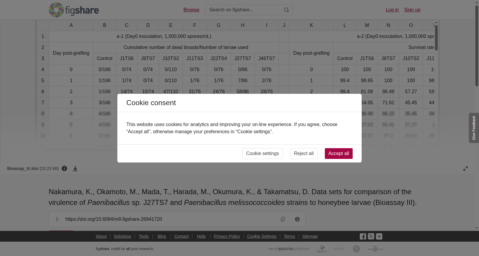
Data sets (cumulative number of dead broods/number of larvae used [a-1, b-1, c-1, and d-1], survival rate [%] [a-2, b-2, c-2, and d-2], larval weight [e], and bacterial load [CFU/mg larva] [f]) for evaluation of the virulence of <i>Paenibacillus</i> sp. J27TS7 and <i>Paenibacillus</i> <i>melissococcoides </i>J1TS6, J6TS7, J10TS2, J11TS3, J22TS4, and J46TS7 to honeybee larvae.The virulence was evaluated by feeding spores of these <i>Paenibacillus</i> strains to <i>in vitro</i>-reared<i> Apis mellifera</i> larvae. For preparation of the spores, bacteria grown on BHI agar (J27TS7) and Columbia<b> </b>blood agar containing 5% sheep blood (CSA) (the other strains) were suspended in saline, and appropriately diluted bacterial suspensions were spread onto BHI agar (J27TS7), MYPGP agar (J6TS7), and CSA (the other strains) plates. The plates were incubated at 35°C for 11–16 days under air plus 5% CO<sub>2</sub> (J6TS7) and aerobic (the other strains) conditions. After sporulation of more than 90% cells was confirmed by staining with malachite green, spores were collected from 10 or more agar plates, suspended in cold sterile H<sub>2</sub>O, and washed four times with cold sterile H<sub>2</sub>O by collecting spores via centrifugation (12,000 x g, 15 min, 4°C), discarding the supernatant, and suspending the spore pellet in 30 mL of cold sterile H<sub>2</sub>O. Washed spores were suspended in 10 mL of cold sterile H<sub>2</sub>O and stored at 4°C. One week or more later, the spores were washed several more times with 30 mL of cold sterile H<sub>2</sub>O to remove organic matter from the lysed vegetative cells. The re-washed spores were suspended in 5–20 mL of cold sterile H<sub>2</sub>O and stored at 4°C until use.Clinically healthy<i> A. mellifera</i> colonies maintained in the Research Institute for Animal Science in Biochemistry and Toxicology, Sagamihara, Japan, were used in this bioassay. From the colonies, less than 24-h-old larvae were grafted onto royal jelly in sterile Petri dishes using a grafting tool and randomly divided into test groups. Each larva was reared in grafting cells placed in a 48-well cell culture plate. The culture plates were kept in a desiccator (relative humidity, 95% ± 5%) and incubated at 34 ± 0.5ºC until day 6 post-grafting (pg). Larvae were infected by feeding them a diet containing spores on day 0 pg or day 2 pg. Inoculum dose (i.e., concentration of spores in the diet) under each condition is shown in or on the top of each table. Larvae that received the inoculum on day 0 pg were transferred to wells with a bacteria-free diet 24 h later, and larvae that received the inoculum on day 2 pg were reared in the same wells thereafter. Larvae in the non-infected control group were fed artificial diets containing the same components as the inocula except the spores. On and after day 7 pg, larvae in the culture plates were incubated in a desiccator at 34 ± 0.5ºC (relative humidity, 80% ± 5%). On day 14 pg, each plate was transferred into an emergence box in a desiccator and incubated at 34 ± 0.5ºC (relative humidity, 80% ± 5%) until day 21 pg.In this bioassay, two groups of larvae were tested for each strain. One was used to assess survival, and the other was used to measure larval weight and bacterial load and/or to analyse histopathologically. After measuring the weights, larvae were stored in a deep freezer at -80°C (for measuring bacterial load) or rinsed for several seconds with sterilised distilled water and fixed in 10% phosphate-buffered formalin (for histopathological analysis). Bacterial loads in the infected larvae were calculated by plating serial dilutions of larval homogenates onto CSA plates and counting colonies on the plates after incubation of the plates at 35ºC for seven days under aerobic conditions. Histopathological analysis results are not included in the data sets. In this bioassay, larval survival was monitored until day 21 pg.
本数据集包含用于评估类芽孢杆菌属(*Paenibacillus*)菌株J27TS7以及蜜蜂类芽孢杆菌(*Paenibacillus melissococcoides*)J1TS6、J6TS7、J10TS2、J11TS3、J22TS4、J46TS7对蜜蜂幼虫毒力的相关检测数据,具体包括:累计死亡幼虫数/所用受试幼虫数[a-1、b-1、c-1、d-1]、存活率(%)[a-2、b-2、c-2、d-2]、幼虫体重[e]以及细菌载量[菌落形成单位(CFU)/毫克幼虫][f]。
本实验通过将上述类芽孢杆菌菌株的孢子喂食体外饲养的西方蜜蜂(*Apis mellifera*)幼虫,以评估其毒力。
关于孢子的制备:将在脑心浸液琼脂(BHI agar)平板上培养的J27TS7菌株,以及在含5%羊血的哥伦比亚血琼脂(Columbia blood agar, CSA)平板上培养的其余菌株,分别悬浮于生理盐水中;将适当稀释的菌液分别涂布于BHI琼脂(J27TS7)、MYPGP琼脂(J6TS7)以及CSA(其余菌株)平板。将平板置于35℃环境下培养:J6TS7菌株需在空气加5% CO₂的条件下培养11~16天,其余菌株则需在有氧环境下培养。通过孔雀绿染色确认超过90%的细胞完成芽孢形成后,从10个及以上的琼脂平板中收集芽孢,悬浮于冷无菌去离子水中;随后通过离心(12000×g,15分钟,4℃)收集芽孢、弃去上清液,将芽孢沉淀重悬于30 mL冷无菌去离子水中,以此方式重复洗涤4次。将洗涤后的芽孢重悬于10 mL冷无菌去离子水中,于4℃条件下保存。一周及以上后,再次用30 mL冷无菌去离子水多次洗涤芽孢,以去除裂解的营养细胞所释放的有机物。重新洗涤后的芽孢重悬于5~20 mL冷无菌去离子水中,于4℃保存至使用。
本生物测定所用的临床健康西方蜜蜂蜂群饲养于日本相模原市的生物化学与毒理学动物科学研究所(Research Institute for Animal Science in Biochemistry and Toxicology)。从该蜂群中选取日龄小于24小时的幼虫,使用移虫针转移至无菌培养皿中的蜂王浆内,并随机分为若干实验组。每头幼虫饲养于置于48孔细胞培养板的移虫杯内。将培养板置于相对湿度为95%±5%的干燥器中,于34±0.5℃下培养至移虫后第6天(pg)。于移虫后第0天或第2天,向幼虫喂食含芽孢的饲料以完成感染。各条件下的接种剂量(即饲料中芽孢的浓度)详见各表格或表格表头。于移虫后第0天接受接种的幼虫在24小时后转移至不含细菌的饲料孔中,而于移虫后第2天接受接种的幼虫则在后续饲养中保留于原孔中。非感染对照组幼虫喂食除不含芽孢外其余成分与接种饲料完全一致的人工饲料。自移虫后第7天起,将培养板置于相对湿度为80%±5%的干燥器中,于34±0.5℃下培养。于移虫后第14天,将每个培养板转移至置于干燥器内的羽化盒中,于34±0.5℃、相对湿度80%±5%的条件下培养至移虫后第21天。
本生物测定中,每个菌株设置两组幼虫:一组用于评估存活率,另一组用于测量幼虫体重、细菌载量或开展组织病理学分析。称量体重后,幼虫要么置于-80℃深冰箱中保存(用于细菌载量检测),要么用无菌蒸馏水冲洗数秒后固定于10%磷酸缓冲福尔马林溶液中(用于组织病理学分析)。感染幼虫的细菌载量通过将幼虫匀浆的系列稀释液涂布于CSA平板上,将平板置于35℃有氧环境下培养7天后计数菌落数计算得到。本数据集未包含组织病理学分析结果。本生物测定中,幼虫存活率的监测持续至移虫后第21天。
提供机构:
figshare创建时间:
2024-09-05
搜集汇总
数据集介绍
以上内容由遇见数据集搜集并总结生成


