
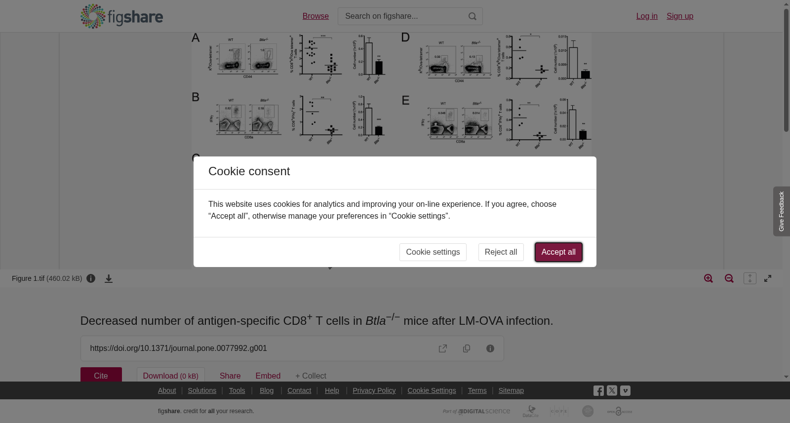
Decreased number of antigen-specific CD8<sup>+</sup> T cells in <i>Btla</i><sup>−/−</sup> mice after LM-OVA infection.
收藏NIAID Data Ecosystem2026-03-07 收录
下载链接:
https://figshare.com/articles/dataset/_Decreased_number_of_antigen_specific_CD8_T_cells_in_Btla_8722_8722_mice_after_LM_OVA_infection_/836840
下载链接
链接失效反馈官方服务:
资源简介:
A- WT and Btla−/− mice were orally infected with 1×10⁹ LM-OVA and 7 days p.i., OVA-specific CD8+ T cells were detected by staining spleen cells with OVA-tetramer (Kb/OVA257–264). The percentage and absolute number of OVA-tetramer+ CD8+ T cells found in WT (circles and open bars) and Btla−/− (squares and filled bars) mice are shown. Each symbol represents an individual mouse from a total of 11–14 mice per group, pooled from three independent experiments. B- IFNγ production by CD8+ T cells isolated from WT and Btla−/− mice, 7 days p.i., was calculated by intracellular staining after ex vivo stimulation of the cells with OVA257–264 peptide. The percentage and absolute number of IFNγ+CD8+ T cells is shown. Data shown correspond to the average of 4–6 mice per group and are representative of three independent experiments. C- OVA-tetramer staining in MLN cells and IEL isolated from LM-OVA infected animals, 7 days p.i. Data pooled from three independent experiments are shown. D- Reduced number of memory CD8+ T cells in Btla−/− mice. The percentage and absolute number of OVA-specific memory CD8+ T cells in the spleen of LM-OVA-infected animals was calculated by staining cells with OVA-tetramers, 70 days p.i. E- IFNγ intracellular staining performed on CD8+ T cells isolated from infected mice 70 days p.i., restimulated ex vivo with OVA257–264 peptide. Data shown correspond to the average of 4 mice per group and are representative of three independent experiments.
A- 野生型(WT)与Btla基因敲除(Btla−/−)小鼠经口感染1×10^9个LM-OVA,于感染后7天(p.i., post infection),通过OVA四聚体(OVA-tetramer,Kb/OVA257–264)染色脾脏细胞以检测OVA特异性CD8+ T细胞。本部分展示了WT组(圆形与空白条)与Btla−/−组(方形与填充条)小鼠中OVA-tetramer阳性(OVA-tetramer+)CD8+ T细胞的百分比与绝对计数。每组共11~14只个体小鼠,数据合并自3次独立实验,每个符号代表单只小鼠。
B- 感染后7天,从WT和Btla−/−小鼠中分离的CD8+ T细胞经OVA257–264肽体外刺激(ex vivo stimulation)后,通过细胞内染色检测干扰素γ(IFNγ)的产生水平。本部分展示了IFNγ阳性CD8+ T细胞(IFNγ+CD8+ T)的百分比与绝对计数。该数据为每组4~6只小鼠的平均值,是3次独立实验的代表性结果。
C- 感染后7天,对LM-OVA感染小鼠的肠系膜淋巴结(mesenteric lymph node, MLN)细胞与肠上皮淋巴细胞(intestinal epithelial lymphocyte, IEL)进行OVA四聚体染色,本部分展示合并自3次独立实验的数据。
D- Btla−/−小鼠的记忆性CD8+ T细胞数量减少。感染后70天,通过OVA四聚体染色检测LM-OVA感染小鼠脾脏中OVA特异性记忆性CD8+ T细胞的百分比与绝对计数。
E- 感染后70天,分离感染小鼠的CD8+ T细胞,经OVA257–264肽体外复刺激(ex vivo restimulation)后进行干扰素γ(IFNγ)细胞内染色。该数据为每组4只小鼠的平均值,是3次独立实验的代表性结果。
创建时间:
2016-02-23
搜集汇总
数据集介绍
以上内容由遇见数据集搜集并总结生成


