CellHIST-Bench
收藏资源简介:
CellHIST-Bench 是一个用于基于组织学的空间基因表达推断的基准数据集。该数据集提供了配对的整张切片组织病理学图像、空间转录组学标签、细胞分割结果、以点为中心的补丁元数据以及细胞与补丁的对应信息。数据集支持两种空间转录组学注释级别:点分辨率空间转录组学(每个点与局部组织学补丁和点级基因表达向量相关联)和单细胞分辨率空间转录组学(每个细胞与单细胞基因表达向量相关联,并可链接到图像检测到的细胞)。CellHIST-Bench 旨在支持从组织病理学图像预测空间基因表达的弱监督学习方法的基准测试。数据集按组织或癌症类型组织,包含整张切片图像、细胞分割文件、补丁元数据、补丁-细胞关系文件和基因表达标签。数据集支持的任务包括点级和单细胞级基因表达预测、弱监督空间基因表达推断、细胞感知表示学习以及跨分辨率空间转录组学分析。数据集还提供了详细的数据字段说明、加载示例和推荐评估设置。
CellHIST-Bench is a benchmark dataset for histology-based spatial gene expression inference. The dataset provides paired whole-slide histopathology images, spatial transcriptomics labels, cell segmentation results, spot-centered patch metadata, and cell-to-patch correspondence information. The dataset supports two levels of spatial transcriptomics annotation: spot-resolution spatial transcriptomics (each spot is associated with a local histology patch and a spot-level gene expression vector) and single-cell-resolution spatial transcriptomics (each cell is associated with a single-cell gene expression vector and can be linked to image-detected cells). CellHIST-Bench is designed to support benchmarking of weakly supervised learning methods for predicting spatial gene expression from histopathology images. The dataset is organized by tissue or cancer type and includes whole-slide images, cell segmentation files, patch metadata, patch-cell relationship files, and gene expression labels. The supported tasks include spot-level and single-cell-level gene expression prediction, weakly supervised spatial gene expression inference, cell-aware representation learning, and cross-resolution spatial transcriptomics analysis. The dataset also provides detailed data field descriptions, loading examples, and recommended evaluation settings.
CellHIST-Bench 数据集概述
数据集简介
CellHIST-Bench 是一个用于组织学空间基因表达推断的基准数据集,提供配对的全切片组织病理学图像、空间转录组标签、细胞分割结果、以斑点为中心的斑块元数据以及细胞与斑块的对应关系信息。
数据集支持两种空间转录组注释级别:
- 斑点分辨率:每个斑点关联一个局部组织学斑块和一个斑点级基因表达向量。
- 单细胞分辨率:每个细胞关联一个单细胞基因表达向量,并可链接到图像检测到的细胞。
数据集组织
数据集按组织或癌症类型组织。每个文件夹包含全切片图像、细胞分割文件、斑块元数据、斑块-细胞关系文件和基因表达标签。
典型目录结构示例:
/data/west_bench ├── LUNG │ ├── wsis │ ├── cellvit++_seg │ ├── patches │ ├── patches_cell │ ├── st_spot_label │ └── st_cell_label ├── IDC ├── SKCM └── ...
数据文件详解
全切片图像(wsis/)
- 文件格式:
.tif - 内容:原始组织病理学图像
- 说明:文件通常很大,需使用 OpenSlide 或 tifffile 等工具读取
细胞分割结果(cellvit++_seg/)
由 CellViT++ 生成,每个样本包含:
| 文件 | 内容 |
|---|---|
*_seg.parquet |
DataFrame,包含 geometry、class、cell_id 等列 |
*.h5 |
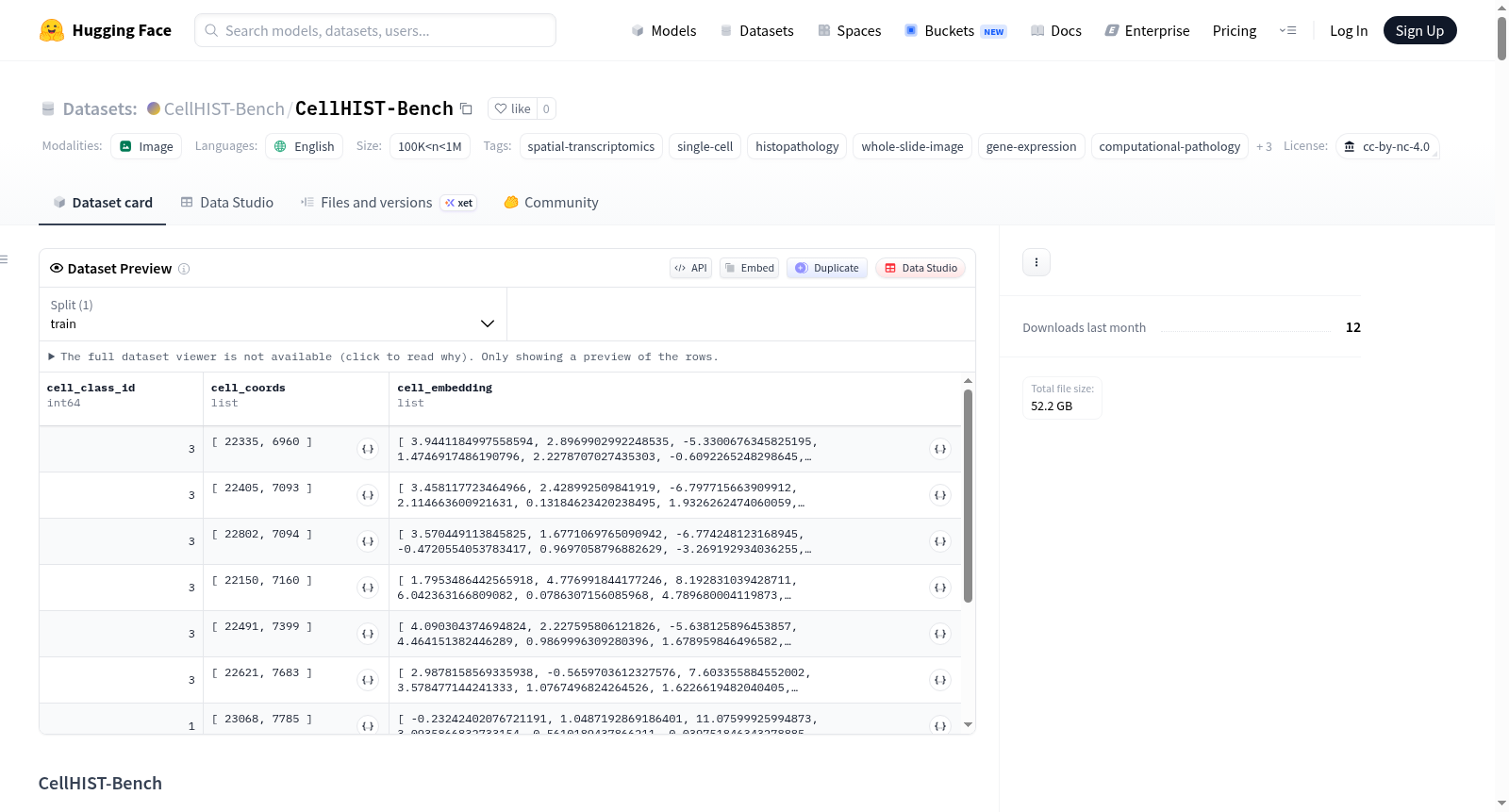
存储 cell_coords(细胞坐标)、cell_embedding(细胞级图像嵌入)、cell_class_id(细胞类别ID) |
斑块元数据(patches/)
- 文件格式:
.h5 - 包含键:
barcode(空间转录组斑点条形码)、coords(斑块在WSI中的坐标) - 注意:不直接存储斑块图像,需根据坐标从对应WSI中裁剪
斑块-细胞关系文件(patches_cell/)
- 文件格式:
.json - 以斑点条形码为索引的字典,每个条目包含:
cell_index:位于斑块中的细胞索引in_spot:指示细胞是否位于斑点区域内
- 每个斑块对应 224×224 像素的局部图像区域
斑点级基因表达标签(st_spot_label/)
- 文件格式:
.h5ad - 包含
num_spots × num_genes的基因表达矩阵 - 行索引对应斑点条形码
单细胞分辨率样本附加文件
| 文件 | 说明 |
|---|---|
*_st_cell_idx.parquet |
空间转录组细胞索引与图像检测细胞ID的映射,包含 st_cell_index 和 cell_id |
st_cell_label/ |
单细胞级基因表达标签,.h5ad 文件,行索引为 st_cell_index |
数据字段汇总
| 文件类型 | 字段 | 说明 |
|---|---|---|
| 全切片图像 | *.tif |
全切片组织病理学图像 |
| 细胞分割 | geometry |
细胞几何或多边形信息 |
class |
预测的细胞类别 | |
cell_id |
WSI中唯一细胞标识符 | |
cell_coords |
细胞坐标 | |
cell_embedding |
细胞级图像嵌入 | |
cell_class_id |
数值型细胞类别ID | |
| 斑块元数据 | barcode |
空间转录组斑点条形码 |
coords |
WSI中斑块坐标 | |
| 斑块-细胞关系 | cell_index |
位于斑块中的细胞索引 |
in_spot |
细胞是否在斑点区域内 | |
| 斑点级标签 | .X |
斑点级基因表达矩阵 |
.obs |
斑点元数据 | |
.var |
基因元数据 | |
| 单细胞级标签 | .X |
单细胞级基因表达矩阵 |
.obs |
细胞元数据 | |
.var |
基因元数据 |
支持的任务
- 斑点级空间基因表达预测:根据斑点中心组织学斑块预测对应的斑点级基因表达向量
- 单细胞级基因表达预测:根据图像衍生细胞信息和局部组织学上下文预测单个细胞的基因表达谱
- 弱监督空间基因表达推断:评估弱监督模型从组织病理学图像推断分子谱的能力
- 细胞感知表示学习:利用细胞分割、细胞嵌入和斑块-细胞关系学习细胞感知的组织学表示
- 跨分辨率空间转录组分析:在统一数据组织下比较斑点级和单细胞级空间基因表达预测
建议的评估设置
斑点级预测
- 输入:斑点中心组织学斑块
- 输出:斑点级基因表达向量
- 标签文件:
st_spot_label/*.h5ad
单细胞级预测
- 输入:细胞级视觉信息和局部组织学上下文
- 输出:单细胞基因表达向量
- 标签文件:
st_cell_label/*.h5ad - 映射文件:
cellvit++_seg/*_st_cell_idx.parquet
数据集划分
数据集可按组织类型、样本ID或任务设置划分。典型划分包括训练集、验证集和测试集。评估泛化能力时,应确保同一生物样本的切片不跨训练集和测试集共享。
预期用途
- 基于组织学的空间基因表达预测
- 空间转录组学的弱监督学习
- 计算病理学
- 细胞感知的组织学表示学习
- 组织学图像与空间组学数据的整合
- 单细胞和空间转录组分析
仅限研究用途。
限制与注意事项
- WSI文件巨大:需专业工具和充足存储
- 斑块图像未直接存储:需用户从WSI中裁剪
- 细胞分割依赖CellViT++:下游分析可能受分割模型准确性影响
- 分辨率差异:斑点和单细胞分辨率样本标签粒度不同,结果需谨慎解读
- 基因表达稀疏性:数据稀疏且含噪声,预测性能需结合生物学和技术因素解释
许可协议
- 许可:CC BY-NC 4.0(知识共享-非商业性使用 4.0 国际许可协议)