3D printed metal structures with inherent micro pores to enhance biodegradability and absorbability
收藏DataCite Commons2025-04-27 更新2025-04-16 收录
下载链接:
https://www.scidb.cn/detail?dataSetId=f60a9504038042e09292136f1206ee31
下载链接
链接失效反馈官方服务:
资源简介:
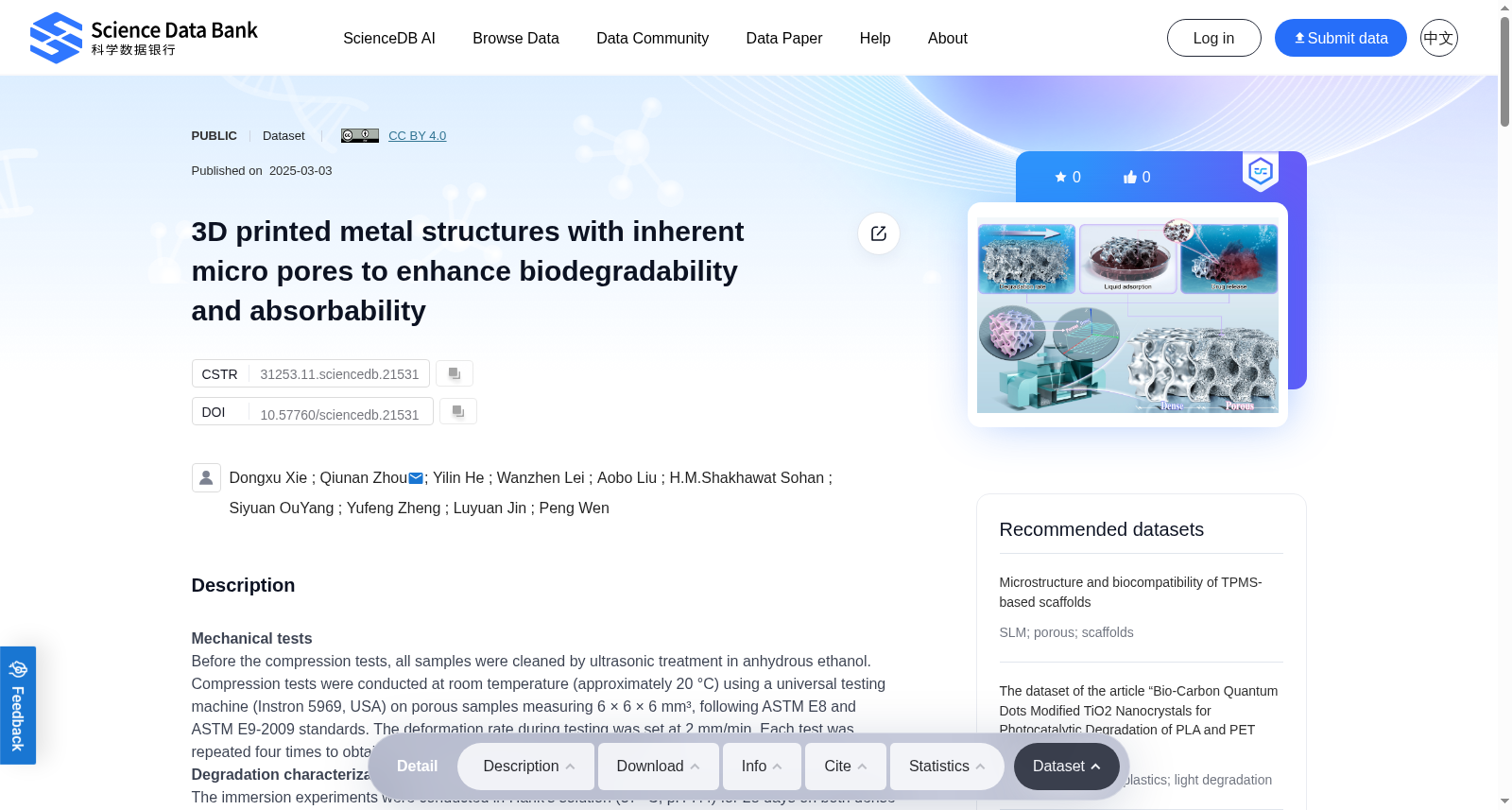
Mechanical testsBefore the compression tests, all samples were cleaned by ultrasonic treatment in anhydrous ethanol. Compression tests were conducted at room temperature (approximately 20 °C) using a universal testing machine (Instron 5969, USA) on porous samples measuring 6 × 6 × 6 mm³, following ASTM E8 and ASTM E9-2009 standards. The deformation rate during testing was set at 2 mm/min. Each test was repeated four times to obtain the average value and standard deviation.Degradation characterizationsThe immersion experiments were conducted in Hank’s solution (37 °C, pH 7.4) for 28 days on both dense and porous samples, following the ASTM G31-72 standard. The dimensions of the porous samples were ϕ10 × 2 mm³ and 6 × 6 × 6 mm³. The volume of Hank’s solution was maintained at a ratio of 20 mL per cm² of sample surface area. The pH of the solution was monitored using a Mettler Five Easy pH meter (FE20 K, Switzerland). After immersion, the samples were rinsed with deionized water. The cleaned porous samples underwent compression testing to investigate the effect of corrosion on mechanical strength. The corroded surfaces were examined using a SEM and EDS. After removing corrosion products with CrO₃ solution, the weight loss of the samples was measured using an electronic balance with an accuracy of ±0.1 mg, and the corrosion rate (CR) was calculated. Each type of test was conducted on four samples to obtain the average value and standard deviation.Electrochemical experimentsThe electrochemical tests were conducted at 37 °C using an electrochemical workstation (Autolab, Metrohm, Switzerland) in Hank’s solution. A standard three-electrode system was employed, with the sample as the working electrode, a platinum plate as the counter electrode, and a saturated calomel electrode as the reference electrode. The tests included open circuit potential (OCP) measurement, electrochemical impedance spectroscopy (EIS) scanning, and potentiodynamic polarization (PDP) testing.The OCP test lasted for 3600 seconds. Following the OCP measurement, EIS was performed over a frequency range of 10⁻² to 10⁵ Hz, with a perturbation voltage of 10 mV. The equivalent circuit was fitted using NOVA 1.11 software (Autolab, Metrohm, Switzerland). The PDP scan was conducted at a scanning rate of 1 mV/s. The corrosion potential (Ecorr) and corrosion current density (icorr) were analyzed using the Tafel extrapolation method and linear fitting. Each test was repeated three times to obtain the average value and standard deviation.Liquid absorption testThe liquid absorption experiments involved immersing implant samples printed with different scan space in a 50% v/v solution of monascus red dye solution. This was done by observing the absorption properties of the porous structures at the bottoms of the samples, which included letters, crowns, spires, and the logo of Tsinghua University printed with a 300 μm scan space. The experiments also measured the time required for the dye to be absorbed to the top of the implant samples with varying Hs to calculate the liquid absorption rate, thereby assessing the effect of different Hs on absorption performance. Each experiment was independently repeated three times to obtain average values and standard deviations.Drug release testIn the drug release experiments, cubes were immersed in drug solutions (monascus red dye solution or minocycline hydrochloride) using a vacuum impregnation method to ensure thorough penetration of the drugs into the micro porous. To slow down drug release, chitosan hydrogel was used to seal the inherent micro pores. Subsequently, the cubes or implants loaded with liquid drugs were immersed in pure water. At predetermined intervals, 500 μL of the release medium was collected and replaced with an equal volume of pure water. The absorbance at specific wavelengths was measured, and the release of the drug over time was monitored according to the calculated standard curve.Cell cultureRat bone marrow mesenchymal stem cells (BMSCs, CP-R131) were obtained from Procell Life Science Co., Ltd. in Wuhan, China. The BMSCs were cultured in α-MEM medium supplemented with 10% FBS (30067334, Gibco, New York, USA) and 1% antibiotics (15070063, Gibco, New York, USA). The medium was completely changed every two days. When the cell confluence reached 90%, the cells were passaged.Direct contact in vitro biological studiesAfter sterilizing the TPMS-structured samples through UV irradiation, they were soaked in m-SBF solution for 48 hours, after which the solution was removed. Each group of samples was then added to culture medium containing 1 × 10^6 cells and incubated for 48 hours. Following incubation, the supernatant was discarded, and the samples were washed twice with PBS. After thorough washing with PBS, the samples underwent gradient dehydration using different concentrations of alcohol (50%, 60%, 70%, 80%, 90%, 95%, and 100%). Finally, the samples were placed in a 24-well plate to dehydrate overnight, and after gold coating, scanning electron microscopy (SEM) was used to assess cell adhesion in each group.Indirect contact in vitro biological studiesIn this study, extract solutions for each group were prepared according to the international standard ISO 10993-5. Samples were immersed in complete culture medium at a ratio of 1.25 ml/cm² of material surface area to culture medium volume and incubated at 37°C for 24 ± 0.5 hours. The solutions were then filtered using a 0.22 μm filter and stored at 4°C. The resulting extracts were diluted tenfold with complete culture medium for subsequent in vitro cell experiments.Cell viability was assessed using the CCK-8 assay and live/dead cell staining. BMSCs were seeded in a 96-well plate at a density of 5000 cells/well. Once the cells adhered, they were treated with complete culture medium containing different extract solutions. The CCK-8 reagent (C0038, Beyotime, Shanghai, China) was prepared according to the manufacturer's instructions. At 24, 48, and 72 hours, the original culture medium was removed, and CCK-8 working solution was added. The cells were incubated for an additional 2 hours before the absorbance at 450 nm was measured using a microplate reader.BMSCs were also seeded in a 12-well plate at a density of 1 × 10^6 cells/well and cultured for 24 hours with the respective media. Live/dead staining working solution was prepared in a ratio of 1 ml: 3 μL: 5 μL (PBS: Calcein AM (Invitrogen): Propidium Iodide (PI, Invitrogen)) and 200 μL of this solution was added to each well. The cells were incubated at 37°C in the dark for 30 minutes before observation under a laser microscope.To induce osteogenic differentiation, BMSCs were seeded in a 6-well plate and, after cell adhesion, cultured in osteogenic induction medium (RAXMX-90021, Saiye, Guangzhou, China) containing the respective composite material extracts. After 7 days of induction, alkaline phosphatase (ALP) staining (P0321S, Beyotime, Shanghai, China) was performed. Additionally, cells were lysed using Western and IP cell lysis buffer without inhibitors (P0013J, Beyotime, Shanghai, China) to extract total protein. The total protein concentration was determined using the BCA protein assay kit (23227, Thermo Fisher, Massachusetts, USA), and ALP activity was quantified using the ALP/AKP assay kit (A059-2-2, Nanjing Jianchen, Nanjing, China). After 14 days of osteogenic induction, Alizarin Red S staining (ARS) (A5533, Sigma, Massachusetts, USA) was conducted. Calcium nodules were then dissolved using 10% hexadecyltrimethylammonium bromide, and the absorbance of each group was measured at 570 nm.
提供机构:
Science Data Bank
创建时间:
2025-03-03
搜集汇总
数据集介绍
背景与挑战
背景概述
该数据集聚焦于3D打印的微孔金属结构,通过机械测试、降解表征、电化学实验、液体吸收测试、药物释放测试和体外生物研究,全面评估了其力学性能、生物降解性、吸收性和生物相容性。所有实验均遵循标准方法并进行了重复测量,数据可靠,适用于生物医学材料研发和优化。
以上内容由遇见数据集搜集并总结生成


