Fundus-AVSeg
收藏资源简介:
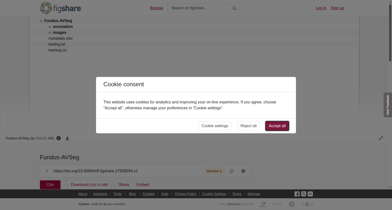
AbstractRetinal artery-vein vessels are associated with systemic chronic diseases and cardiovascular diseases. Therefore, the accurate quantitative analysis of retinal artery-vein vessels is the preliminary basis of clinical diagnosis. Most of the existing artificial intelligence(AI) methods are data-driven. Although some public retinal artery-vein vessel segmentation datasets have been released, their data quality is unsatisfactory. In this paper, we establish a new fundus image dataset for AI-based artery-vein segmentation, Fundus-AVSeg. It consists of 100 high-resolution fundus images with pixel-wise manual annotation by professional ophthalmologists. We believe our Fundus-AVSeg will benefit the further development of retinal artery-vein vessel segmentation.<br>Data Information Fundus-AVSeg contains 100 fundus images, 40 of which are from normal fundus, 20 from diabetic retinopathy(DR) patients, 20 from age-related macular degeneration (AMD), and 20 from glaucoma. Images have two resolutions: 2656×1992 and 1280×1280. Pixel-wise manual annotated categories include arteries, veins, artery-vein crossings, and vessels of uncertain category. Two image quality categories are also provided for each image: low-quality and high-quality. Data CollectionThe data source of Fundus-AVSeg is Shenzhen Eye Hospital. All 100 fundus images are obtained from the imaging database of Shenzhen Eye Hospital. The fundus images are captured by ophthalmologists using ZEISS VISUCAM200 fundus cameras or Canon fundus cameras, which are the mainstream products of fundus cameras. All fundus images are generated during real clinical diagnostic processes. Approval of all ethical and experimental procedures and protocols is granted by the Shenzhen Eye Hospital under the ETHICAL NUMBER 2022KYPJ062. Data RecordsThe Fundus-AVSeg dataset has been uploaded to Figshare in the form of a zipped file. The unzipped file contains two folders and one Microsoft Office Excel list, and two txt format files, named ``images'', ``annotation'' , ``metadata.xlsx'', ``training.txt'', and ``testing.txt'', respectively. In the ``images'' folder, there are 100 fundus images. Images are named ``n\_D/A/G/N.png", where ``n'' means the number of fundus images and ``D'', ``A'', ``G'', and ``N'' stand for ``DR'', ``AMD'', ``Glaucoma'', and ``Normal''. The ``annotation'' folder contains 100 corresponding annotated images, which are named according to the same rule, where a specific image in this folder is the ground truth of the image with the same name in the ``images''. The ``metadata.xlsx'' is an Excel file that holds the following information: image name, eye ID, disease type, and image quality. The ``training.txt'' and ``testing.txt'' files store the specific image names for training and testing, respectively, following an 8:2 split of the dataset. Please note that the current data split strategy is recommended by us and can be altered for different research purposes.Usage NotesThe complete dataset is available for download via the provided link. Users have the flexibility to divide the dataset based on their specific study designs. It is expected that users will reference this paper in their research and recognize the dataset’s contribution to their studies.Code AvailabilityThe code mentioned in this study can be found at https://github.com/AI-thpremed/Basic-Seg-Experiment.<br><br>
视网膜动静脉血管与全身性慢性疾病及心血管疾病密切相关。因此,对视网膜动静脉血管进行精准的定量分析是临床诊断的初步基础。当前多数人工智能(AI)方法均为数据驱动型。尽管已有部分公开的视网膜动静脉血管分割数据集发布,但这些数据集的数据质量不尽如人意。本文构建了一款面向AI动静脉分割任务的新型眼底图像数据集——Fundus-AVSeg。该数据集包含100张高分辨率眼底图像,且由专业眼科医生逐像素进行手动标注。我们相信,Fundus-AVSeg将助力视网膜动静脉血管分割领域的进一步发展。 数据集概况 Fundus-AVSeg共包含100张眼底图像,其中40张来自正常眼底,20张来自糖尿病视网膜病变(DR)患者,20张来自年龄相关性黄斑变性(AMD)患者,剩余20张来自青光眼患者。图像包含两种分辨率:2656×1992与1280×1280。逐像素手动标注的类别包括动脉、静脉、动静脉交叉区以及类别不确定的血管。此外,每张图像还被标注为两种图像质量等级之一:低质量与高质量。 数据采集 Fundus-AVSeg的数据来源为深圳眼科医院。所有100张眼底图像均取自深圳眼科医院的影像数据库,由眼科医生使用蔡司(ZEISS)VISUCAM200眼底相机或佳能(Canon)眼底相机拍摄——这两款均为当前主流的眼底相机产品。所有眼底图像均采集自真实的临床诊断流程。本研究的所有伦理与实验流程均已获得深圳眼科医院批准,伦理编号为2022KYPJ062。 数据记录 Fundus-AVSeg数据集已以压缩包形式上传至Figshare平台。解压后的压缩包包含两个文件夹、一个Microsoft Office Excel表格文件以及两个TXT格式文件,分别命名为"images"、"annotation"、"metadata.xlsx"、"training.txt"与"testing.txt"。在"images"文件夹中,存放有100张眼底图像,图像命名格式为"n_D/A/G/N.png",其中"n"代表图像编号,"D"、"A"、"G"、"N"分别代表糖尿病视网膜病变(DR)、年龄相关性黄斑变性(AMD)、青光眼(Glaucoma)以及正常(Normal)。"annotation"文件夹包含100张对应的标注图像,命名规则与图像文件一致,该文件夹内的每张标注图像均与"images"文件夹中同名图像一一对应,作为其真值标签。"metadata.xlsx"为Excel表格文件,存储以下信息:图像名称、眼ID、疾病类型以及图像质量。"training.txt"与"testing.txt"文件分别存储训练集与测试集的具体图像名称,数据集按照8:2的比例划分。需要说明的是,本次推荐的数据划分策略仅供参考,用户可根据不同研究需求进行调整。 使用须知 完整数据集可通过指定链接下载。用户可根据自身研究设计灵活划分数据集。同时,期望使用者在研究中引用本文,并认可本数据集对其研究的贡献。 代码获取 本研究提及的代码可在https://github.com/AI-thpremed/Basic-Seg-Experiment获取。