
Angelou0516/PROMISE12
收藏Hugging Face2026-04-30 更新2026-05-03 收录
下载链接:
https://hf-mirror.com/datasets/Angelou0516/PROMISE12
下载链接
链接失效反馈官方服务:
资源简介:
---
license: other
license_name: attribution
tags:
- medical-imaging
- segmentation
- prostate
- mri
- multi-center
task_categories:
- image-segmentation
pretty_name: 'PROMISE12: Prostate MR Image Segmentation 2012'
configs:
- config_name: default
data_files:
- split: train
path: data/train-*
dataset_info:
features:
- name: case_id
dtype: string
- name: split
dtype: string
- name: num_slices
dtype: int32
- name: height
dtype: int32
- name: width
dtype: int32
- name: image
dtype: image
- name: mask
dtype: image
- name: overlay
dtype: image
splits:
- name: train
num_bytes: 10219680
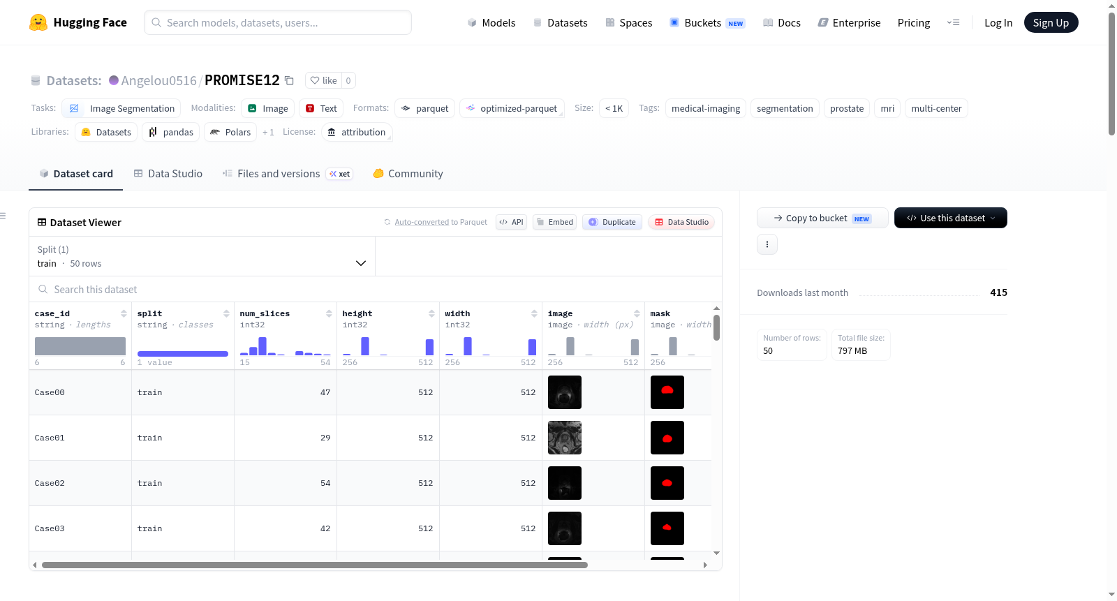
num_examples: 50
download_size: 10227947
dataset_size: 10219680
---
# PROMISE12: Prostate MR Image Segmentation 2012
Transverse T2-weighted prostate MRI volumes from the MICCAI 2012 Grand Challenge for whole-gland prostate segmentation. Contributions from 4 clinical centers.
## Dataset Details
| Property | Value |
|---|---|
| **Modality** | MRI (T2-weighted, transverse) |
| **Organ** | Prostate (whole gland) |
| **Total cases** | 50 (training only — see below) |
| **Format** | MetaImage (`.mhd` header + `.raw` binary) |
| **Image dtype** | int16 (`MET_SHORT`) |
| **Mask dtype** | int8 (`MET_CHAR`), binary {0, 1} |
| **In-plane size** | 512 × 512 |
| **Slices per volume** | 15 – 54 |
## Scope of This Upload
The original PROMISE12 challenge released 100 cases:
- **50 Training** — public images **and** ground-truth masks (this upload)
- **30 Test** — images released, ground-truth was withheld (challenge-closed, not included here)
- **20 Live Challenge** — images released, ground-truth was withheld (not included here)
Only the 50 training cases have publicly available masks, so this repository contains those 50 paired volumes. The original challenge is closed and online scoring of test/live predictions is no longer available.
## Multi-Center Composition
Each contributing center provided 25 of the 100 original cases. The 50 training cases here are a mix from these centers:
| Center | Institution | Field | Endorectal Coil | Vendor |
|---|---|---|---|---|
| RUNMC | Radboud Univ. Nijmegen Medical Centre, NL | 3T | No | Siemens |
| BIDMC | Beth Israel Deaconess Medical Center, USA | 3T | Yes | GE |
| UCL | University College London, UK | 1.5T & 3T | No | Siemens |
| HK | Haukeland Univ. Hospital, Norway | 1.5T | Yes | Siemens |
Note: The BIDMC contribution here is a distinct subset from the `Angelou0516/BIDMC` dataset (which is the FedDG Multi-Site Prostate MRI collection containing 12 BIDMC cases).
## File Structure
Each case has 4 files (image + mask, each MetaImage `.mhd` header + `.raw` binary):
```
CaseXX.mhd # T2 MRI volume header (references CaseXX.raw)
CaseXX.raw # T2 MRI voxel data (int16)
CaseXX_segmentation.mhd # Prostate mask header (references CaseXX_segmentation.raw)
CaseXX_segmentation.raw # Binary prostate mask (uint8, {0, 1})
```
50 cases total: `Case00` through `Case49`.
## Ground Truth
Masks were contoured slice-by-slice by an experienced reader at each contributing center using 3DSlicer or MeVisLab (spline-connected points), then independently validated and corrected by a second expert reviewer. This is the only mask source publicly distributed.
A second-observer mask (used in the original paper to establish inter-observer variability) exists for the test/live cases but is not part of this release.
## Loading Example
```python
import SimpleITK as sitk
image = sitk.GetArrayFromImage(sitk.ReadImage("Case00.mhd")) # (Z, Y, X) int16
mask = sitk.GetArrayFromImage(sitk.ReadImage("Case00_segmentation.mhd")) # (Z, Y, X) uint8
print(image.shape, mask.shape)
```
## License
`Other (Attribution)` — see `LICENSE.TXT`. Citation of the paper below is mandatory.
## Citation
```bibtex
@article{litjens2014promise12,
title = {Evaluation of prostate segmentation algorithms for {MRI}: the {PROMISE12} challenge},
author = {Litjens, Geert and Toth, Robert and van de Ven, Wendy and Hoeks, Caroline and
Kerkstra, Sjoerd and van Ginneken, Bram and Vincent, Graham and Guillard, Gwenael and
Birbeck, Neil and Zhang, Jindang and Strand, Robin and Malmberg, Filip and Ou, Yangming and
Davatzikos, Christos and Kirschner, Matthias and Jung, Florian and Yuan, Jing and
Qiu, Wu and Gao, Qiang and Edwards, Philip Eddie and Maan, Bianca and
van der Heijden, Ferdinand and Ghose, Soumya and Mitra, Jhimli and Dowling, Jason and
Barratt, Dean and Huisman, Henkjan and Madabhushi, Anant},
journal = {Medical Image Analysis},
volume = {18},
number = {2},
pages = {359--373},
year = {2014},
doi = {10.1016/j.media.2013.12.002}
}
```
## Sources
- Zenodo mirror: https://zenodo.org/records/8014041 (DOI: 10.5281/zenodo.8014041)
- Original portal: https://promise12.grand-challenge.org/
PROMISE12 is a medical imaging dataset for prostate MRI image segmentation, originating from the MICCAI 2012 Grand Challenge. The dataset includes 50 training cases, each comprising T2-weighted MRI images and corresponding prostate segmentation masks. Data is sourced from four different clinical centers, featuring a multi-center composition. The dataset is in MetaImage format, consisting of .mhd header files and .raw binary files. Image data is of int16 type, while masks are of int8 type with binary values of 0 or 1. Each case has an image size of 512×512, with the number of slices ranging from 15 to 54. Ground truth was contoured slice-by-slice by an experienced reader and independently validated by a second expert reviewer. The dataset is suitable for image segmentation tasks, particularly focusing on whole-gland prostate segmentation.
提供机构:
Angelou0516
搜集汇总
数据集介绍
构建方式
PROMISE12数据集源自2012年MICCAI前列腺MR图像分割大挑战,汇聚了来自四个临床中心的T2加权横向前列腺MRI容积图像。原始挑战共发布100例病例,其中仅有50例训练数据同时公开了影像与对应的金标准分割掩膜。影像数据以MetaImage格式存储,包含.mhd头文件与.raw二进制体素数据,图像维度为512×512,切片数量介于15至54层之间。掩膜由各中心的资深阅片者利用3DSlicer或MeVisLab软件逐层勾画,并经过第二位专家独立验证与修正,确保了标注的高质量与一致性。
特点
该数据集具有鲜明的多中心、多场强及多厂商特性,其病例来自荷兰、美国、英国和挪威的四家顶级医学研究机构,涵盖了3T与1.5T两种磁场强度,并涉及有无直肠内线圈的采集方案以及Siemens与GE等不同厂商设备。这一异质性构建了真实临床场景下的数据分布,使得PROMISE12成为评估前列腺分割算法泛化能力的标杆性基准。数据仅包含50例训练配对的体素级数据,既提供了丰富的解剖变异,又因其规模有限而有效考验模型在小样本多中心数据上的鲁棒性。
使用方法
使用PROMISE12数据集时,研究者需通过SimpleITK库高效加载MetaImage格式的原始数据:调用sitk.ReadImage读取.mhd文件后以sitk.GetArrayFromImage获取形状为(Z, Y, X)的影像与掩膜数组,影像为int16类型,掩膜为uint8类型的二值图像。数据以病例为单位组织,命名格式为Case00至Case49。该版本仅包含训练子集,原始挑战的测试集与实时挑战集因金标准未公开而无法使用。建议在训练中沿用标准5折交叉验证策略以充分利用有限样本,并严格遵循论文引用以尊重原作者贡献。
背景与挑战
背景概述
PROMISE12(Prostate MR Image Segmentation 2012)数据集源自2012年MICCAI国际会议举办的前列腺MRI分割挑战赛,由Radboud大学医学中心等四家临床机构联合创建,核心研究团队包括Geert Litjens等学者。该数据集专注于解决前列腺磁共振图像中全腺体的分割问题,包含50例训练样本(公开图像与金标准掩膜),覆盖不同场强、线圈类型及扫描设备的多中心数据,为评估和比较分割算法提供了标准化基准。自2014年发表以来,PROMISE12已成为医学图像分割领域的里程碑式数据集,其发表的论文《Evaluation of prostate segmentation algorithms for MRI: the PROMISE12 challenge》被广泛引用,极大推动了前列腺MRI分割技术的研究进展,对放射组学、计算机辅助诊断及临床治疗规划具有深远影响。
当前挑战
PROMISE12数据集所解决的领域挑战在于前列腺全腺体的精确分割,MRI图像中前列腺边界模糊、灰度分布不均匀,且受不同成像设备、场强及有无内插线圈等多中心差异影响,导致传统算法泛化能力不足。构建过程中,数据集面临多中心数据标注的高昂成本与一致性难题,需由各中心经验丰富的读片者逐层勾画并由第二专家独立验证校正,以确保金标准掩膜的可靠性。此外,原始挑战赛仅公开50例训练样本的标注,测试集与在线挑战集的掩膜未开放,限制了后续算法的全面评估与公平比较,使得模型在跨中心场景下的泛化性能验证仍是一个待解决的挑战。
常用场景
经典使用场景
PROMISE12数据集作为前列腺MRI分割领域的标杆基准,广泛应用于深度学习模型的性能评估与算法对比。研究者通常利用该数据集的全腺体分割任务,训练并验证U-Net、nnU-Net等经典架构,亦或探索基于Transformer或扩散模型的前沿方法。数据集中来自四个临床中心的50例T2加权横断面MRI图像,涵盖了不同场强、是否使用直肠内线圈等多种成像条件,为评估分割算法在多中心异构数据上的泛化能力提供了标准测试平台。其清晰的任务定义和公开的标注掩膜,使其成为前列腺分割研究中最常引用的公共资源之一。
解决学术问题
该数据集针对的核心学术问题是前列腺MRI图像中全腺体边界的精确自动分割。在临床实践中,手动勾画前列腺轮廓耗时且易受操作者间差异影响,而传统半自动方法在低对比度区域或腺体形态变异时表现不稳。PROMISE12通过提供多中心、多供应商、多场强的高质量标注数据,系统性推动了计算机辅助分割算法的发展。它帮助研究者量化了不同算法在异质性数据上的分割精度,揭示了成像参数(如梯度强度、线圈类型)对分割性能的影响,从而促进了更具鲁棒性和泛化能力的模型设计,显著提升了自动化前列腺分割的临床可信度。
衍生相关工作
PROMISE12作为奠基性数据集,催生了大量经典工作。2014年的挑战赛原始论文汇总了多种传统方法,标志着该领域的首次全面比较。此后,U-Net及其变体(如Attention U-Net、nnU-Net)在此数据上验证了优越性,其中nnU-Net通过自动化配置流程屡次刷新性能记录。在域适应方向,PROMISE12常与BIDMC、RUNMC等其他数据集联合使用,用于测试无监督或半监督域适应算法,以解决不同医疗中心间的图像分布偏移问题。此外,一些利用伪标签或对比学习来缓解标注稀疏性的半监督方法,也常以该数据集作为评估基准,验证其在不完全监督环境下的有效性。
以上内容由遇见数据集搜集并总结生成



