BreCaHAD: A Dataset for Breast Cancer Histopathological Annotation and Diagnosis
收藏DataCite Commons2025-06-01 更新2024-07-27 收录
下载链接:
https://figshare.com/articles/BreCaHAD_A_Dataset_for_Breast_Cancer_Histopathological_Annotation_and_Diagnosis/7379186/3
下载链接
链接失效反馈官方服务:
资源简介:
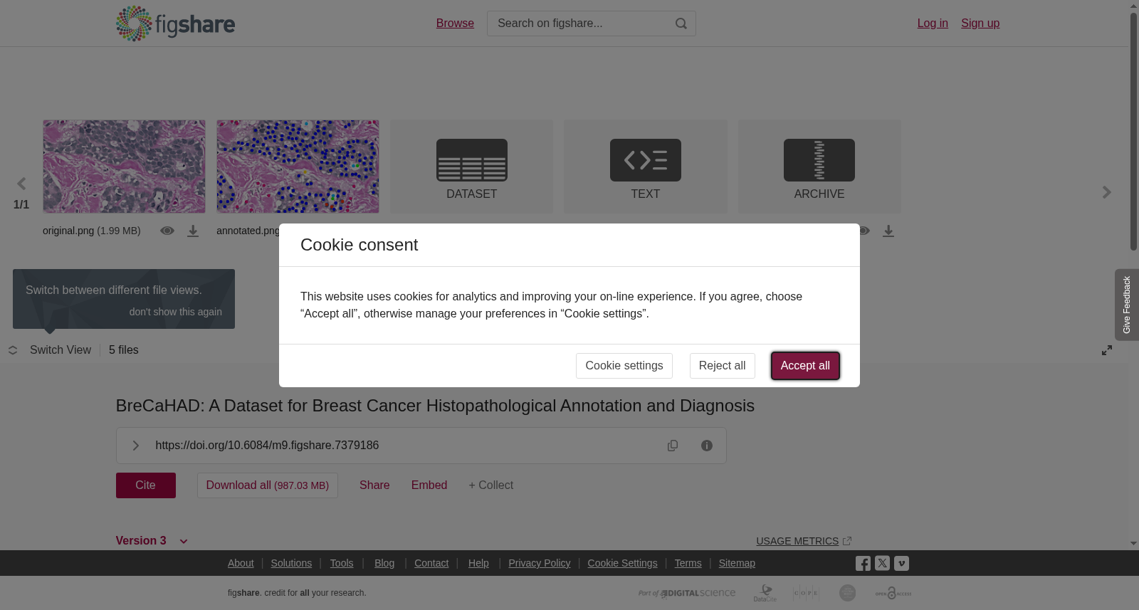
This dataset consists of 1 <b>.xlsx</b> file, 2 <b>.png</b> files, 1 <b>.json</b> file and 1 <b>.zip</b> file:<br><b>annotation_details.xlsx: </b>The distribution of annotations in the previously mentioned six classes (mitosis, apoptosis, tumor nuclei, non-tumor nuclei, tubule, and non-tubule) is presented in a Excel spreadsheet.<br><b>original.png: </b>The input image.<br><br><b>annotated.png:</b> An example from the dataset. In the annotated image, blue circles indicate the tumor nuclei, pink circles show non-tumor nuclei such as blood cells, stroma nuclei, and lymphocytes; orange and green circles are mitosis and apoptosis, respectively; light blue circles are true lumen for tubules, and yellow circles represent white regions (non-lumen) such as fat, blood vessel, and broken tissues.<br><br><b>data.json:</b> The annotations for the BreCaHAD dataset are provided in JSON (JavaScript Object Notation) format. In the given example, the JSON file (ground truth) contains two mitosis and only one tumor nuclei annotations. Here, <i>x</i> and <i>y</i> are the coordinates of the centroid of the annotated object, and the values are between [0, 1] (divided by width and height of an image).<br><br><b>BreCaHAD.zip: </b>An archive file containing dataset. Three folders are included: images (original images), groundTruth (json files), and groundTruth_display (groundTruth applied on original images)<br><br>
本数据集包含1个<b>.xlsx</b>文件、2个<b>.png</b>文件、1个<b>.json</b>文件以及1个<b>.zip</b>文件:<br><b>annotation_details.xlsx:</b>该Excel表格展示了前文提及的六类标注(有丝分裂(mitosis)、细胞凋亡(apoptosis)、肿瘤细胞核(tumor nuclei)、非肿瘤细胞核(non-tumor nuclei)、小管(tubule)与非小管(non-tubule))的分布情况。<br><b>original.png:</b>输入图像。<br><br><b>annotated.png:</b>数据集中的示例图像。在该标注图像中,蓝色圆圈代表肿瘤细胞核(tumor nuclei),粉色圆圈表示非肿瘤细胞核(non-tumor nuclei,如血细胞、基质细胞核与淋巴细胞);橙色圆圈与绿色圆圈分别对应有丝分裂(mitosis)与细胞凋亡(apoptosis);浅蓝色圆圈代表小管的真实管腔(true lumen),黄色圆圈则代表脂肪、血管与破损组织等非管腔区域(non-lumen)。<br><br><b>data.json:</b>BreCaHAD数据集的标注以JSON(JavaScript Object Notation)格式提供。在本示例中,该JSON文件(即真值标注(ground truth))包含2个有丝分裂标注与1个肿瘤细胞核标注。此处的<i>x</i>与<i>y</i>为标注对象的质心坐标,其取值范围为[0, 1](通过图像的宽高进行归一化得到)。<br><br><b>BreCaHAD.zip:</b>包含完整数据集的归档文件,内含3个文件夹:images(原始图像文件夹)、groundTruth(真值标注JSON文件文件夹)与groundTruth_display(将真值标注叠加至原始图像后的展示文件夹)。
提供机构:
figshare
创建时间:
2019-01-28
搜集汇总
数据集介绍
以上内容由遇见数据集搜集并总结生成


