HCP-MMP1.0 volumetric (NIfTI) masks in native structural space
收藏Mendeley Data2024-01-31 更新2024-06-28 收录
下载链接:
https://figshare.com/articles/dataset/HCP-MMP1_0_volumetric_NIfTI_masks_in_native_structural_space/4249400/5
下载链接
链接失效反馈官方服务:
资源简介:
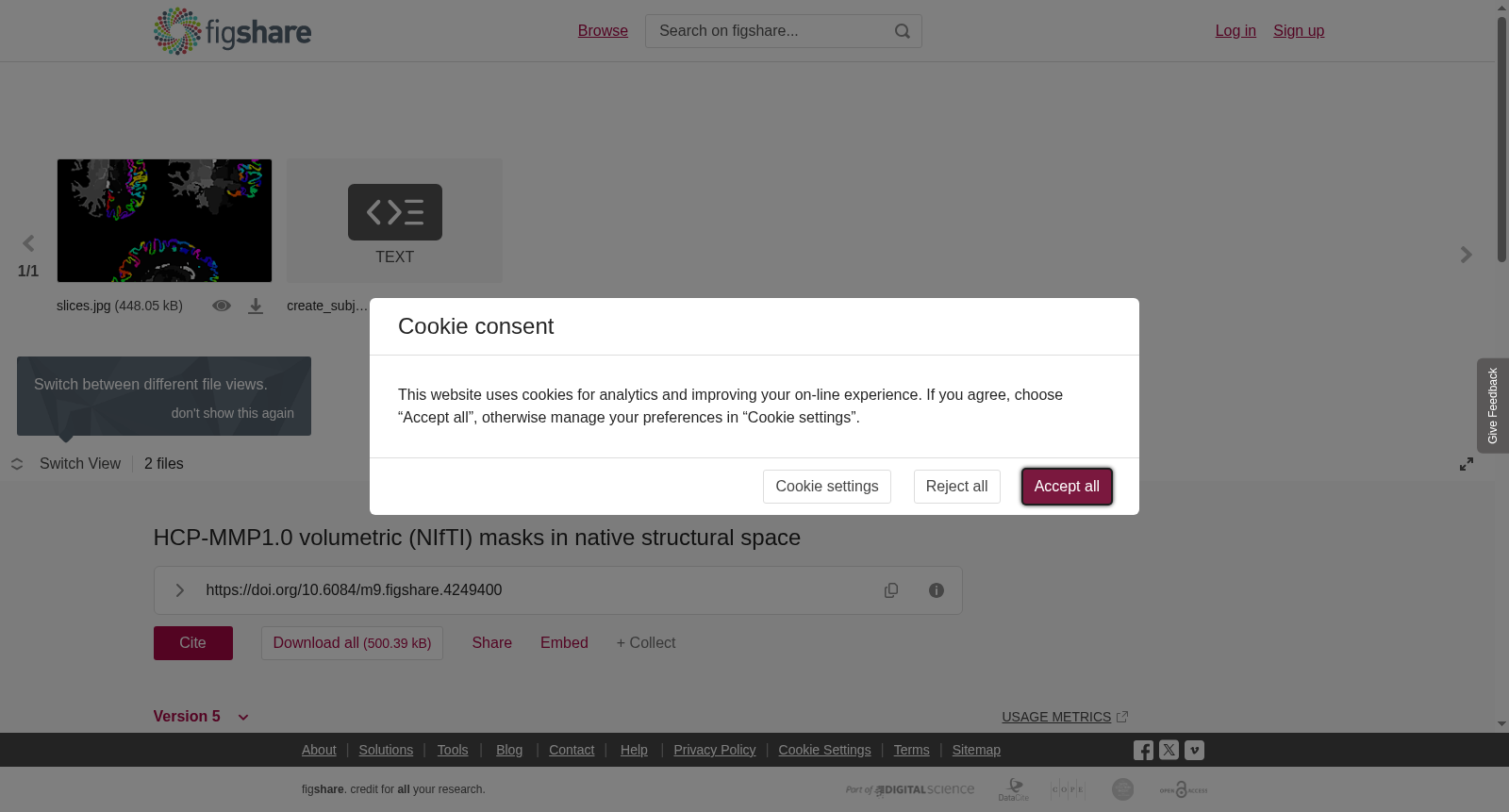
We in our group received with great interest the publication of the HCP-MMP 1.0 parcellation by Glasser et al. (Nature) created using data from the Human Connectome Project earlier this year. Often in our connectivity pipelines we use volume files for parcellation in native space, so we decided to try and convert the Connectome Workbench files to volume masks in native structural space to try out in future studies. We were happy to find that someone had already gone through the trouble of generating FreeSurfer annotation files projected on fsaverage, so all we had to do was find a way to transform these annot files to each subject’s space and convert them to volume masks. So we wrote this Linux shell script that performs a series of conversion and transformation steps using only FreeSurfer commands. It first converts the annotation files (lh.HCPMMP1.annot and rh.HCPMMP1.annot, downloaded from https://figshare.com/articles/HCP-MMP1_0_projected_on_fsaverage/3498446) to labels using mri_annotation2label, then takes each label from fsaverage to each subject’s space with mri_label2label, converts transformed labels back to annotation with mri_label2annot, and finally converts these to volume files (nii.gz) with mris_label2annot. Seems like too many steps, but this is how we (who are not FreeSurfer experts) got satisfactory results. The default final file consists of a single .nii.gz volume containing the cortical HCP-MMP1.0 regions plus the subcortical regions from the FreeSurfer segmentation, each region assigned a unique voxel value. It should be noted that the HCP-MMP1.0 parcellation includes 180 regions per hemisphere - 179 cortical and one subcortical (hippocampus). In the final volume file, left-hemisphere cortical HCP-MMP1.0 regions will have values between 1001 and 1181, whereas right-sided regions will have values between 2001 and 2181. The correspondence between each specific region and its voxel value is given in a look-up table that is saved in each subject’s output folder. To identify the hippocampi (and other subcortical structures), one needs to check the corresponding voxel value given in the FreeSurferColorLUT.txt file provided with FreeSurfer (https://surfer.nmr.mgh.harvard.edu/fswiki/FsTutorial/AnatomicalROI/FreeSurferColorLUT), as they are generated based on the original aseg parcellation*. *In the previous version of the script, a few perihippocampal cortical voxels were ascribed values (1121 and 2121) that should correspond to the hippocampus in the HCPMMP1.0 parcellation. Since only the cortical regions from this parcellation are generated, these voxels are now assigned the values corresponding to the hippocampus as defined by the automatic FreeSurfer subcortical segmentation (17 and 53). Optionally, one can choose to also generate individual volume files for each cortical and/or subcortical parcellation region. This option requires FSL. If the user chooses to create individual subcortical masks, the FreeSurferColorLUT.txt must also be available in the base ($SUBJECTS_DIR/) folder. By default, the script also generates tables with anatomical information for each cortical region (number of vertices, area, volume, mean thickness, etc.). Instructions on how to use the script can be found in the script itself, or here:https://cjneurolab.org/2016/11/22/hcp-mmp1-0-volumetric-nifti-masks-in-native-structural-space/ Hope this can be of use!
本团队近期对Glasser等人于《Nature》发表的、基于人类连接组计划(Human Connectome Project)数据构建的HCP-MMP 1.0脑区分割图谱产生了浓厚兴趣。我们在常规连接组分析流程中,常使用原生空间下的体积文件进行脑区分割,因此计划将连接组工作台(Connectome Workbench)格式的文件转换为原生结构空间下的体积掩码,以供后续研究使用。
我们欣喜地发现,已有研究者完成了映射至fsaverage模板的FreeSurfer注释(annotation)文件的生成工作,因此我们仅需将这些annot文件转换至每个被试的原生空间,并进一步转换为体积掩码即可。为此,我们编写了一款仅使用FreeSurfer命令完成一系列转换与配准步骤的Linux Shell脚本。
该脚本首先通过mri_annotation2label工具,将从https://figshare.com/articles/HCP-MMP1_0_projected_on_fsaverage/3498446下载的注释文件lh.HCPMMP1.annot与rh.HCPMMP1.annot转换为标签文件;随后利用mri_label2label工具,将fsaverage模板下的每个标签配准至各被试的原生空间;再通过mri_label2annot工具将配准后的标签重新转换为注释文件;最终借助mris_label2annot工具将其转换为体积文件(.nii.gz格式)。尽管步骤略显繁琐,但作为非FreeSurfer专业使用者的我们,通过该流程获得了令人满意的结果。
默认输出的最终文件为单个.nii.gz体积文件,其中包含皮层层面的HCP-MMP1.0脑区,以及FreeSurfer分割得到的皮层下脑区,每个脑区对应唯一的体素值。需说明的是,HCP-MMP1.0分割方案每个半球包含180个脑区:179个皮层脑区与1个皮层下脑区(海马体)。在最终体积文件中,左侧半球皮层HCP-MMP1.0脑区的体素值范围为1001至1181,右侧半球对应脑区的体素值范围则为2001至2181。各具体脑区与其体素值的对应关系,将以查找表的形式保存于每个被试的输出文件夹中。
若需识别海马体(及其他皮层下结构),需参考FreeSurfer配套提供的FreeSurferColorLUT.txt文件(https://surfer.nmr.mgh.harvard.edu/fswiki/FsTutorial/AnatomicalROI/FreeSurferColorLUT)中给出的对应体素值,因为这些结构是基于原始aseg分割方案生成的*。
*在该脚本的旧版本中,少量海马旁皮层体素被赋予了体素值1121与2121,这两个值本应对应HCP-MMP1.0分割方案中的海马体。但由于本脚本仅生成该分割方案中的皮层脑区,因此现已将这些体素的取值修改为FreeSurfer自动皮层下分割定义的海马体对应值:17与53。
用户可选择为每个皮层和/或皮层下分割脑区生成独立的体积掩码文件,该功能依赖FSL工具包。若用户需要生成独立的皮层下掩码文件,则需确保FreeSurferColorLUT.txt文件存在于根目录($SUBJECTS_DIR/)下。默认情况下,脚本还会为每个皮层脑区生成包含解剖学信息的表格,内容包括顶点数、表面积、体积、平均皮层厚度等。
该脚本的使用说明可参见脚本内部注释,或访问以下链接:https://cjneurolab.org/2016/11/22/hcp-mmp1-0-volumetric-nifti-masks-in-native-structural-space/。希望本工具能对您有所帮助!
创建时间:
2024-01-31
搜集汇总
数据集介绍
背景与挑战
背景概述
该数据集提供了HCP-MMP1.0分区在原生结构空间中的体积(NIfTI)掩码,通过一个Linux shell脚本实现从FreeSurfer注释文件到体积掩码的转换。最终文件包含皮层和皮层下区域,每个区域有唯一的体素值,适用于神经科学和连接组学研究。
以上内容由遇见数据集搜集并总结生成


