
CCEP ECoG dataset across age 4-51
收藏OpenNeuro2022-07-21 更新2026-03-14 收录
下载链接:
https://openneuro.org/datasets/ds004080
下载链接
链接失效反馈官方服务:
资源简介:
# Dataset description
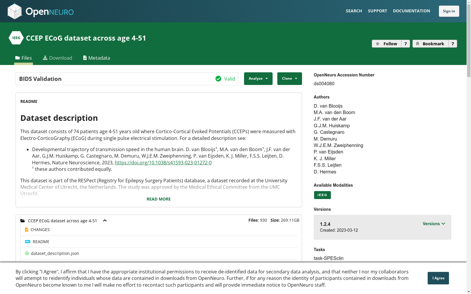
This dataset consists of 74 patients age 4-51 years old where Cortico-Cortical Evoked Potentials (CCEPs) were measured with Electro-CorticoGraphy (ECoG) during single pulse electrical stimulation. For a detailed description see:
- Developmental trajectory of transmission speed in the human brain. D. van Blooijs¹, M.A. van den Boom¹, J.F. van der Aar, G.J.M. Huiskamp, G. Castegnaro, M. Demuru, W.J.E.M. Zweiphenning, P. van Eijsden, K. J. Miller, F.S.S. Leijten, D. Hermes, Nature Neuroscience, 2023, https://doi.org/10.1038/s41593-023-01272-0
¹ these authors contributed equally.
This dataset is part of the RESPect (Registry for Epilepsy Surgery Patients) database, a dataset recorded at the University Medical Center of Utrecht, the Netherlands. The study was approved by the Medical Ethical Committee from the UMC Utrecht.
## Contact
- Dorien van Blooijs: D.vanBlooijs@umcutrecht.nl
- Frans Leijten: F.S.S.leijten@umcutrecht.nl
- Dora Hermes: hermes.dora@mayo.edu
# Data organization
This data is organized according to the Brain Imaging Data Structure specification. A community-driven specification for organizing neurophysiology data along with its metadata. For more information on this data specification, see https://bids-specification.readthedocs.io/en/stable/
Each patient has their own folder (e.g., `sub-ccepAgeUMCU01` to `sub-ccepAgeUMCU74`) which contains the iEEG recordings data for that patient, as well as the metadata needed to understand the raw data and event timing.
Data are logically grouped in the same BIDS session and stored across runs that indicating the day and time point of recording during the monitoring period.
If extra electrodes were added/removed during this period, the session was divided into different sessions (e.g. ses-1a and ses-1b).
We use the optional run key-value pair to specify the day and the start time of the recording (e.g. run-021315, day 2 after implantation, which is day 1 of the monitoring period, at 13:15).
The task key-value pair in long-term iEEG recordings describes the patient's state during the recording of this file. The task label is “SPESclin“ since these files contain data collected during clinical single pulse electrical stimulation (SPES).
Electrode positions include Destrieux atlas labels that were estimated by running Freesurfer on the individual subject MRI scan and taking the most common surface label within a sphere around the electrode. All shared electrode positions were then converted to MNI152 space using the Freesurfer surface based non-linear transformation. We note that this surface based transformation distorts the dimensions of the grids, but maintains the gyral anatomy.
# License
This dataset is made available under the Public Domain Dedication and License CC v1.0, whose full text can be found at
https://creativecommons.org/publicdomain/zero/1.0/.
We hope that all users will follow the ODC Attribution/Share-Alike Community Norms (http://www.opendatacommons.org/norms/odc-by-sa/);
in particular, while not legally required, we hope that all users of the data will acknowledge by citing the following in any publication:
Developmental trajectory of transmission speed in the human brain, D. van Blooijs, M.A. van den Boom, J.F. van der Aar, G.J.M. Huiskamp, G. Castegnaro, M. Demuru, W.J.E.M. Zweiphenning, P. van Eijsden, K. J. Miller, F.S.S. Leijten, D. Hermes, Nature Neuroscience, 2023, https://doi.org/10.1038/s41593-023-01272-0
# Code
Code to analyses these data is available at: https://github.com/MultimodalNeuroimagingLab/mnl_ccepAge
# Acknowledgements
We thank the SEIN-UMCU RESPect database group (C.J.J. van Asch, L. van de Berg, S. Blok, M.D. Bourez, K.P.J. Braun, J.W. Dankbaar, C.H. Ferrier, T.A. Gebbink, P.H. Gosselaar, R. van Griethuysen, M.G.G. Hobbelink, F.W.A. Hoefnagels, N.E.C. van Klink, M.A. van ‘t Klooster, G.A.P. deKort, M.H.M. Mantione, A. Muhlebner, J.M. Ophorst, P.C. van Rijen, S.M.A. van der Salm, E.V. Schaft, M.M.J. van Schooneveld, H. Smeding, D. Sun, A. Velders, M.J.E. van Zandvoort, G.J.M. Zijlmans, E. Zuidhoek and J. Zwemmer) for their contributions and help in collecting the data, and G. Ojeda Valencia for proofreading the manuscript.
# Funding
Research reported in this publication was supported by the National Institute of Mental Health of the National Institutes of Health under Award Number R01MH122258 (DH, FSSL, the content is solely the responsibility of the authors and does not necessarily represent the official views of the National Institutes of Health), the EpilepsieNL under Award Number NEF17-07 (DvB) and the UMC Utrecht Alexandre Suerman MD/PhD Stipendium 2015 (WZ).
# 数据集说明
本数据集包含74名年龄介于4至51岁的患者,在单脉冲电刺激期间,通过皮层脑电图(Electro-CorticoGraphy, ECoG)记录了皮层-皮层诱发电位(Cortico-Cortical Evoked Potentials, CCEPs)。详细描述请参见:
- 《人脑内传导速度的发育轨迹》,D. van Blooijs¹、M.A. van den Boom、J.F. van der Aar、G.J.M. Huiskamp、G. Castegnaro、M. Demuru、W.J.E.M. Zweiphenning、P. van Eijsden、K. J. Miller、F.S.S. Leijten、D. Hermes,《自然-神经科学》,2023,https://doi.org/10.1038/s41593-023-01272-0
¹ 上述作者贡献均等。
本数据集隶属于RESPect(癫痫手术患者登记库)数据库,该数据集采集自荷兰乌得勒支大学医学中心。本研究已通过乌得勒支大学医学中心医学伦理委员会审批。
## 联系方式
- 多里安·范·布卢伊斯:D.vanBlooijs@umcutrecht.nl
- 弗朗斯·S.S. 莱滕:F.S.S.leijten@umcutrecht.nl
- 多拉·赫姆斯:hermes.dora@mayo.edu
# 数据组织
本数据遵循脑成像数据结构(Brain Imaging Data Structure, BIDS)规范进行组织——这是一项由社区推动的、用于整理神经生理学数据及其元数据的规范。有关该数据规范的更多信息,请参阅:https://bids-specification.readthedocs.io/en/stable/
每位患者拥有独立的文件夹(例如`sub-ccepAgeUMCU01`至`sub-ccepAgeUMCU74`),其中包含该患者的颅内脑电图(intracranial electroencephalogram, iEEG)录制数据,以及用于理解原始数据与事件时序的元数据。
数据在逻辑上归属于同一个BIDS会话,并存储于多个序列(run)中,序列标识对应监测期间的录制日期与时间点。若在此期间新增/移除了额外电极,则将会话拆分为不同子会话(例如ses-1a与ses-1b)。
我们使用可选的run键值对来指定录制日期与开始时间(例如run-021315表示植入术后第2天,即监测期第1天,13:15开始录制)。
长期颅内脑电图录制中的task键值对用于描述该文件录制期间患者的状态。本次任务标签为“SPESclin”,因为这些文件包含临床单脉冲电刺激(single pulse electrical stimulation, SPES)期间采集的数据。
电极位置包含Destrieux图谱(Destrieux Atlas)标注结果:该标注是通过对个体受试者的磁共振成像扫描运行FreeSurfer软件,并取电极周围球体内最常见的皮层表面标注得到的。随后,所有共享的电极位置均通过基于FreeSurfer表面的非线性变换转换至MNI152空间(MNI152 Space)。需注意,该基于表面的变换会使电极网格的维度产生畸变,但可保留脑回解剖结构。
# 许可协议
本数据集采用公共领域贡献与许可协议CC v1.0进行授权,完整协议文本可在https://creativecommons.org/publicdomain/zero/1.0/查阅。
我们期望所有使用者遵循ODC署名-相同方式共享社区规范(http://www.opendatacommons.org/norms/odc-by-sa/);具体而言,尽管无法律强制要求,但我们希望所有数据使用者在任何出版物中通过引用以下文献予以致谢:
《人脑内传导速度的发育轨迹》,D. van Blooijs、M.A. van den Boom、J.F. van der Aar、G.J.M. Huiskamp、G. Castegnaro、M. Demuru、W.J.E.M. Zweiphenning、P. van Eijsden、K. J. Miller、F.S.S. Leijten、D. Hermes,《自然-神经科学》,2023,https://doi.org/10.1038/s41593-023-01272-0
# 代码
用于分析本数据集的代码可在以下网址获取:https://github.com/MultimodalNeuroimagingLab/mnl_ccepAge
# 致谢
我们感谢SEIN-UMCU RESPect数据库团队(C.J.J. van Asch、L. van de Berg、S. Blok、M.D. Bourez、K.P.J. Braun、J.W. Dankbaar、C.H. Ferrier、T.A. Gebbink、P.H. Gosselaar、R. van Griethuysen、M.G.G. Hobbelink、F.W.A. Hoefnagels、N.E.C. van Klink、M.A. van ‘t Klooster、G.A.P. deKort、M.H.M. Mantione、A. Muhlebner、J.M. Ophorst、P.C. van Rijen、S.M.A. van der Salm、E.V. Schaft、M.M.J. van Schooneveld、H. Smeding、D. Sun、A. Velders、M.J.E. van Zandvoort、G.J.M. Zijlmans、E. Zuidhoek以及J. Zwemmer)为数据采集提供的贡献与协助,同时感谢G. Ojeda Valencia对本文手稿的校对工作。
# 资助
本出版物所报道的研究得到了美国国立卫生研究院下属国家精神卫生研究所的资助,资助编号为R01MH122258(DH、FSSL,本内容仅代表作者个人观点,不代表美国国立卫生研究院的官方立场)、EpilepsieNL资助编号NEF17-07(DvB)以及乌得勒支大学医学中心2015年Alexandre Suerman医学博士/博士奖学金(WZ)。
创建时间:
2022-07-21
搜集汇总
数据集介绍
背景与挑战
背景概述
该数据集包含74名年龄在4至51岁之间的患者,在单脉冲电刺激期间使用皮层脑电图(ECoG)测量皮层-皮层诱发电位(CCEP),旨在研究人类大脑传输速度的发育轨迹。数据遵循BIDS规范组织,包括iEEG记录、电极位置信息(如Destrieux图谱标签和MNI152空间转换),总大小约为269.11GB,覆盖广泛的年龄范围,支持神经科学研究。
以上内容由遇见数据集搜集并总结生成



