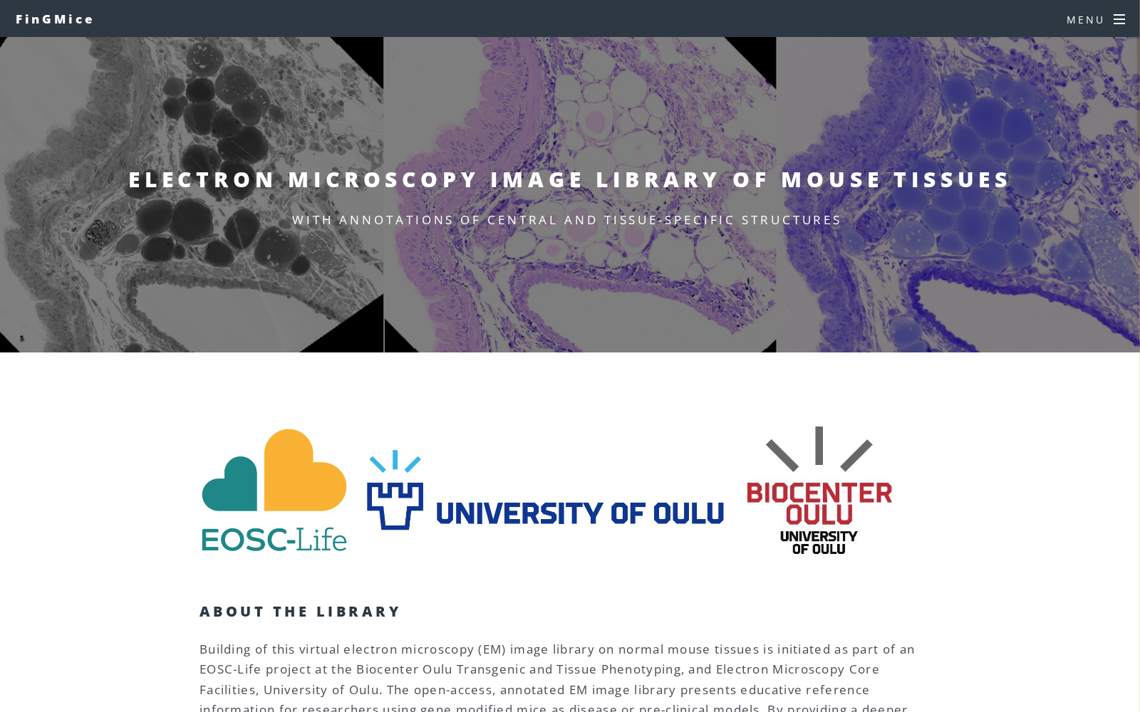
Electron Microscopy Image Library of Mouse Tissues
收藏资源简介:
Building of this virtual electron microscopy (EM) image library on normal mouse tissues is initiated as part of an EOSC-Life project at the Biocenter Oulu Transgenic and Tissue Phenotyping, and Electron Microscopy Core Facilities, University of Oulu, Finland, in collaboration with Infrafrontier. The open-access, annotated EM image library presents educative reference information for researchers using gene modified mice as disease or pre-clinical models. By providing a deeper understanding of tissues at ultrastructural level, it eases and aids in understanding structural changes and related phenotypes in mouse models. This resource is currently in development.
本针对正常小鼠组织的虚拟电子显微镜(EM)图像库的构建工作,系芬兰奥卢大学奥卢生物中心转基因与组织表型及电子显微镜核心设施所承担的EOSC-Life项目的组成部分,本次构建与Infrafrontier合作开展。该开放获取、带注释的电子显微镜图像库,可为以基因修饰小鼠作为疾病模型或临床前模型的研究人员提供教学参考资料。通过帮助研究者从超微结构层面深化对组织的认知,该图像库可辅助理解小鼠模型中的结构变化及其相关表型。目前该资源仍处于开发阶段。