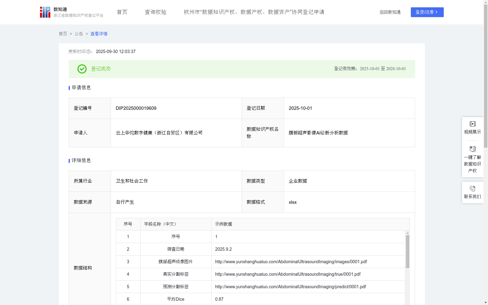
腹部超声影像AI诊断分析数据
收藏浙江省数据知识产权登记平台2025-09-30 更新2025-10-04 收录
下载链接:
https://www.zjip.org.cn/home/announce/trends/187953
下载链接
链接失效反馈官方服务:
资源简介:
一、适用条件与对象
腹部超声影像AI诊断分析数据适用于接入超声设备的基层医院、医学影像AI公司、设备制造商及科研院所。
主要服务对象包括基层医师、研发工程师和科研人员,尤其适合资源有限但需提升诊断精度与效率的基层医院。
二、解决的问题和痛点
(1)有效应对基层医院因医师经验差异导致的诊断一致性低、漏诊误诊率高的问题;
(2)突破传统超声设备智能化水平不足、功能单一的瓶颈;
(3)缓解学术界与工业界因高质量、标准化标注数据缺乏而导致的模型研发与迭代困难。
三、有益效果
(1)为基层医师提供实时病灶识别、特征自动量化与结果判定,显著提升诊断准确性与效率;
(2)为设备厂商提供即插即用的AI模块,增强产品竞争力与市场差异化;
(3)为科研机构提供经过严格质控的腹部超声标准化数据集,助力算法研发与临床转化。
四、外部复用价值
(1)核心算法可授权集成至多品牌超声设备及PACS系统;
(2)脱敏结构化数据库支持多中心研究、模型协同训练与持续优化;
(3)标准化诊断流程与质控体系可推广至基层单位,促进医疗资源下沉与分级诊疗落地。一、数据采集
通过全数字超声诊断仪,采集腹部超声影像图片,收集影像相关的结构化特征信息。
核心字段:
腹部超声影像图片:通过URL存储腹部超声影像图片(URL链接已脱敏)
真实分割标签:专家标注的真实分割标签(精确标注病灶类型)
预测分割标签:模型识别的预测分割标签
腹部超声数据:检查部位、位置、形态、大小、边界、内部特征
诊断数据:AI诊断结果判定、疾病名称、临床意义
辅助字段:筛查日期用于数据管理。
二、数据处理
对所有腹部超声影像图片进行标准化预处理:尺寸归一化(统一超声影像图片分辨率)、图像去噪(减少超声影像斑点噪声)、亮度对比度调整(优化超声影像质量)、特征数据向量化处理
目标:提升后续算法模型的鲁棒性和分割精度
三、核心算法规则 (模型构建与训练)
(1)模型架构:
采用编码器-解码器结构的卷积神经网络(CNN)
模型公式:P=CNNθ(I)
I:预处理后的腹部超声影像图片
CNNθ:卷积神经网络模型参数
P:腹部病灶的预测分割标签的图片
(2)训练策略:
损失函数:结合Dice损失和二元交叉熵损失
优化器:Adam
性能评估指标:平均Dice系数、平均IoU。在训练和验证阶段,使用平均Dice系数和平均IoU作为核心指标,量化模型预测分割标签结果与真实分割标签之间的重叠精度。
(3)多特征融合:
将腹部超声影像特征与临床特征(位置、形态、大小、边界、内部特征)融合,输出AI诊断结果判定、疾病名称与临床意义。
四、数据应用
(1)临床诊断辅助:自动生成腹部病灶分割结果及量化参数,输出AI诊断结果判定和疾病名称;
(2)质量控制:实时监测分割质量(平均Dice≥0.80,平均IoU≥0.70),异常案例自动标记复核;
(3)科研价值:脱敏的腹部超声影像特征数据库支持模型迭代优化,多中心研究数据标准化支持。
1. Application Conditions and Target Users
This abdominal ultrasound imaging AI diagnosis and analysis dataset is applicable to primary hospitals connected to ultrasound equipment, medical imaging AI companies, equipment manufacturers and research institutions. The main target users include primary physicians, R&D engineers and researchers, and it is particularly suitable for primary hospitals with limited resources but in need of improving diagnostic accuracy and efficiency.
2. Solved Problems and Pain Points
(1) Effectively address the problems of low diagnostic consistency and high missed diagnosis and misdiagnosis rates caused by differences in physician experience in primary hospitals;
(2) Break through the bottlenecks of insufficient intelligence level and single functions of traditional ultrasound equipment;
(3) Alleviate the difficulties in model development and iteration faced by academia and industry due to the lack of high-quality, standardized annotated data.
3. Beneficial Effects
(1) Provide real-time lesion recognition, automatic feature quantification and result judgment for primary physicians, significantly improving diagnostic accuracy and efficiency;
(2) Provide plug-and-play AI modules for equipment manufacturers, enhancing product competitiveness and market differentiation;
(3) Provide abdominal ultrasound standardized datasets with strict quality control for research institutions, facilitating algorithm development and clinical translation.
4. External Reusability Value
(1) The core algorithm can be licensed for integration into multi-brand ultrasound equipment and PACS systems;
(2) The de-identified structured database supports multi-center research, model collaborative training and continuous optimization;
(3) The standardized diagnostic process and quality control system can be promoted to primary medical institutions, promoting the downward transfer of medical resources and the implementation of hierarchical diagnosis and treatment.
1. Data Collection
Abdominal ultrasound images are collected via full-digital ultrasound diagnostic equipment, and structured feature information related to the images is collected.
Core fields:
- Abdominal ultrasound image: Stored via URL (the URL links have been de-identified)
- Ground truth segmentation labels: Real segmentation labels annotated by experts (accurately annotating lesion types)
- Predicted segmentation labels: Predicted segmentation labels identified by the model
- Abdominal ultrasound data: Examination site, location, morphology, size, boundary, internal features
- Diagnostic data: AI diagnostic result judgment, disease name, clinical significance
Auxiliary field: Screening date for data management.
2. Data Processing
Standardized preprocessing is performed on all abdominal ultrasound images, including: size normalization (unifying the resolution of ultrasound images), image denoising (reducing speckle noise in ultrasound images), brightness and contrast adjustment (optimizing ultrasound image quality), and vectorization processing of feature data.
Objective: Improve the robustness and segmentation accuracy of subsequent algorithm models.
3. Core Algorithm Rules (Model Construction and Training)
(1) Model Architecture:
A convolutional neural network (CNN) with an encoder-decoder structure is adopted.
Model formula: $P=CNN_ heta(I)$
Where:
$I$: Preprocessed abdominal ultrasound images
$CNN_ heta$: Convolutional neural network model parameters
$P$: Images of predicted segmentation labels of abdominal lesions
(2) Training Strategy:
Loss function: Combining Dice loss and binary cross-entropy loss
Optimizer: Adam
Performance evaluation metrics: Average Dice coefficient, average IoU. In the training and validation stages, the average Dice coefficient and average IoU are used as core metrics to quantify the overlapping accuracy between the model's predicted segmentation label results and the ground truth segmentation labels.
(3) Multi-feature Fusion:
Fuse abdominal ultrasound imaging features with clinical features (location, morphology, size, boundary, internal features) to output AI diagnostic result judgment, disease name and clinical significance.
4. Data Application
(1) Clinical Diagnosis Assistance: Automatically generate abdominal lesion segmentation results and quantitative parameters, and output AI diagnostic result judgment and disease name;
(2) Quality Control: Real-time monitoring of segmentation quality (average Dice ≥ 0.80, average IoU ≥ 0.70), and automatic marking and review of abnormal cases;
(3) Scientific Research Value: The de-identified abdominal ultrasound imaging feature database supports model iterative optimization and provides standardized support for multi-center research data.
提供机构:
云上华佗数字健康(浙江自贸区)有限公司
创建时间:
2025-09-05
搜集汇总
数据集介绍
背景与挑战
背景概述
该数据集包含861条腹部超声影像数据,每日更新,采用xlsx格式存储,涵盖影像图片、分割标签、量化指标(如平均Dice和IoU)及临床特征,用于AI辅助诊断。其核心价值在于通过卷积神经网络算法实现病灶自动分割和诊断判定,有效提升基层医院诊断准确性和效率,并支持设备厂商和科研机构的模型优化与多中心研究。
以上内容由遇见数据集搜集并总结生成


