nih-chest-xray-14
收藏资源简介:
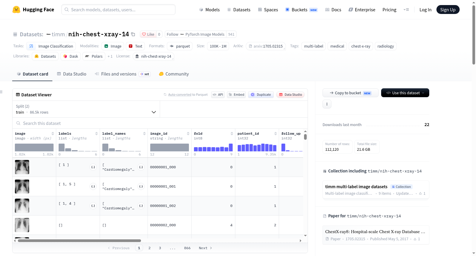
NIH ChestX-ray14数据集(NIH Clinical Center)是CVPR 2017论文中ChestX-ray8的扩展版本,包含112,120张正面胸部X光图像,来自30,805名独特患者。每张图像通过自然语言处理(NLP)从关联的放射学报告中挖掘出14种胸部病理标签(多标签)。14种常见胸部病理包括:Atelectasis、Cardiomegaly、Consolidation、Edema、Effusion、Emphysema、Fibrosis、Hernia、Infiltration、Mass、Nodule、Pleural_Thickening、Pneumonia、Pneumothorax。约54%的图像在源数据中被标记为“No Finding”,这些图像的标签列表为空;注意“No Finding”不等于“正常”,这些图像可能显示14个类别之外的模式或不确定发现。标签通过NLP提取,具有噪声(NIH估计准确率>90%),测试标签同样来自NLP过程,未经放射科医生验证。数据集按患者级别进行划分:训练集86,524张图像(28,008名患者),测试集25,596张图像(2,797名患者),无患者同时出现在两个划分中。训练集包含一个fold列(0-9),基于多标签迭代分层分配(Sechidis等人,2011,种子42),按患者分组,每个患者的所有图像都在同一个fold中,可用于交叉验证。图像为原始的1024x1024 8位PNG,重新编码为8位灰度JPEG(质量95),平均PSNR约48.4 dB,SSIM约0.988,保留全部256个灰度级。每个样本包含字段:image(JPEG图像)、labels(阳性发现的ClassLabel索引序列,空表示No Finding)、label_names(阳性发现名称)、image_id(原始图像文件名)、fold(训练集的fold编号,测试集为null)、patient_id、follow_up、patient_age、patient_sex、view_position(PA/AP)、original_width、original_height、pixel_spacing_x、pixel_spacing_y(来自原始元数据),以及bboxes(边界框列表,仅880张测试图像有,共984个框,覆盖8种发现)。该数据集适用于多标签图像分类任务,标准评估指标是每个发现下的ROC AUC平均值。使用不受限制,但需承认NIH Clinical Center并引用CVPR 2017论文。
The NIH ChestX-ray14 dataset (NIH Clinical Center) is an extended version of ChestX-ray8 from the CVPR 2017 paper, containing 112,120 frontal-view chest X-ray images from 30,805 unique patients. Each image is annotated with up to 14 thoracic pathology labels (multi-label) mined from associated radiology reports using natural language processing (NLP). The 14 common thoracic pathologies include: Atelectasis, Cardiomegaly, Consolidation, Edema, Effusion, Emphysema, Fibrosis, Hernia, Infiltration, Mass, Nodule, Pleural_Thickening, Pneumonia, Pneumothorax. Approximately 54% of images are labeled as No Finding, meaning the label list is empty; note that No Finding does not equal normal, as these images may show patterns outside the 14 categories or uncertain findings. Labels are extracted via NLP and are noisy (NIH estimates accuracy >90%); test labels also come from the NLP process and have not been validated by radiologists. The dataset is split at the patient level: training set 86,524 images (28,008 patients), test set 25,596 images (2,797 patients), with no patient appearing in both splits. The training set includes a fold column (0-9) assigned by multi-label iterative stratification (Sechidis et al., 2011, seed 42), grouped by patient, so all images of a patient are in the same fold, enabling cross-validation. Images are originally 1024x1024 8-bit PNG, re-encoded as 8-bit grayscale JPEG (quality 95), with average PSNR ~48.4 dB, SSIM ~0.988, preserving all 256 gray levels. Each sample contains fields: image (JPEG image), labels (sequence of ClassLabel indices for positive findings, empty for No Finding), label_names (names of positive findings), image_id (original image filename), fold (fold number for training set, null for test set), patient_id, follow_up, patient_age, patient_sex, view_position (PA/AP), original_width, original_height, pixel_spacing_x, pixel_spacing_y (from original metadata), and bboxes (list of bounding boxes, only 880 test images have them, total 984 boxes covering 8 findings). The dataset is suitable for multi-label image classification tasks; the standard evaluation metric is the average ROC AUC across findings. Use is unrestricted, but NIH Clinical Center should be acknowledged and the CVPR 2017 paper cited.